


Actin-driven protrusions generate rapid long-range membrane tension propagation in cells
Preprint posted on 8 September 2022 https://www.biorxiv.org/content/10.1101/2022.09.07.507005v1
Navigating a tense situation: the plasma membrane and actin cortex form an integrated system for rapid long-range tension propagation.
Selected by Nicolaes Hyun-Kee MinCategories: bioengineering, biophysics, cell biology
Background
Cells are surrounded by physical forces, which bend, poke, and stretch the plasma membrane, altering membrane tension1,2. Forces applied from within the cell by membrane-tethered actin protrusions and cortical contraction also modulate membrane tension2. Tension not only governs the shape of cells, but also orchestrates mechanosensitive ion channel opening, receptor trafficking, and endocytosis, which all profoundly affect fundamental cell behaviours like migration, proliferation, and differentiation3-6.
Tension has been observed to rapidly propagate throughout the plasma membrane upon local mechanical stimulation6-9. However, there is compelling evidence that suggests the plasma membrane resists tension propagation10. Currently, it is unclear whether the plasma membrane facilitates or resists tension propagation. Moreover, the relative contribution from the plasma membrane and underlying actin cortex to tension propagation has not been resolved. The authors addressed this by measuring tension propagation upon force inflicted locally on both the membrane and actin cortex using optogenetics and micropipette aspiration, and on the membrane only by optical trap pulling.
Key findings
Local actin protrusion elicits cell-wide membrane tension propagation
The authors sought to establish a system that could elicit local membrane stretch while simultaneously measuring tension at a distant site. Thus, they used Opto-PI3K, an optogenetic approach for inducing highly localized and controlled actin protrusion-driven membrane stretch in neutrophil-like HL-60 cells11,12. Opto-PI3K employs two constructs: (1) membrane-bound iLiD, which is a synthetic protein that undergoes a conformational change upon blue light (488nm) illumination to bind to SspB peptide, and (2) SspB linked to the PI3K iSH2 domain, which constitutively binds endogenous activated PI3K. Upon illumination with blue light, iLiD (1) binds to SspB (2), inducing membrane localization of iSH2 and recruitment of activated PI3K. This drives PIP3 production, downstream Rac activation, and rapid actin protrusion. To measure tension at a distant site, the authors used optical trapping of lectin-coated beads that bind tightly to the membrane. Using this ingenious system (Figure 1 left), they demonstrated that local actin protrusion-driven membrane stretching results in rapid tension propagation to the opposite side of the cell (Figure 1 right), which is in line with previous observations that support a role for the cell membrane in propagating tension6-9.
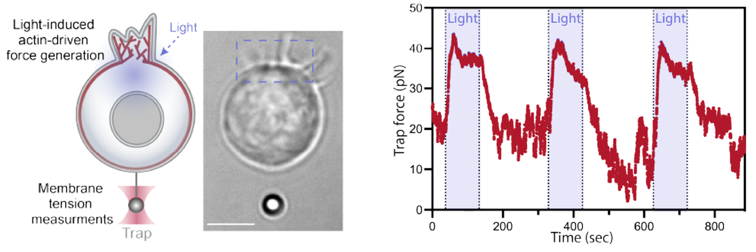
Figure 1. Left: experimental system to simultaneously measure membrane tension while optogenetically inducing actin protrusion. Right: tension increases rapidly upon local actin protrusion on opposite side of the cell.
Forces acting on both plasma membrane and actin cortex drive tension propagation while pulling on membrane alone does not
The authors next aimed to resolve the observed discrepancy of plasma membrane resistance against tension propagation10. In the study from Shi and colleagues10, force was applied to the membrane by optical trapping, pulling only the membrane and not the underlying cytoskeleton. To attempt to reproduce the results from this earlier study, the authors utilized a dual tether system, where one optical trap was moved to apply force and the other was kept static to measure tension (Figure 2 left). If propagation would occur, the force measured on the static trap would increase proportionally with movement of the mobile trap. However, consistent with previous observations, movement of the mobile trap did not increase tension measurement on the static trap (Figure 2 right), indicating that force acting on the plasma membrane alone does not drive tension propagation.
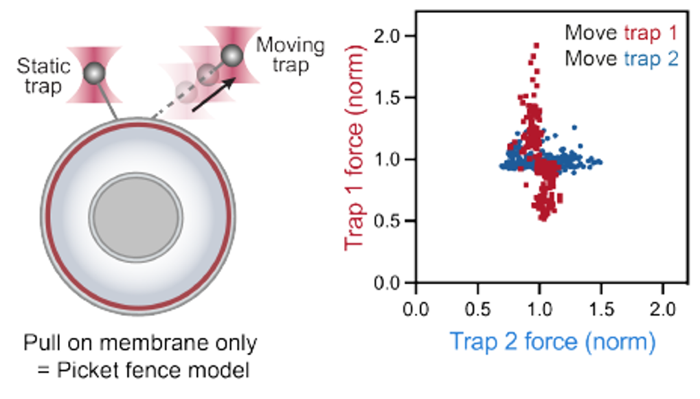
Figure 2. Left: dual tether system to assess tension propagation with force applied to the membrane only. Right: tension measurement on the static trap remains constant with force from the mobile trap.
To explain these observations, the authors devised a composite model made up of the elastic plasma membrane, gel-like actin cortex, and adhesive linkers connecting the two. According to this model, tension acting on the plasma membrane alone is resisted by the actin cortex, whereas tension on the cortex and membrane propagates throughout the cell. Simulating membrane tension using this model reproduced the experimental results, where pulling on the membrane alone did not propagate tension and pulling on the cortex generated rapid tension propagation. To further validate this model, the authors performed micropipette aspiration, where a section of the cell is pulled into a micropipette tip by suction, inflicting pulling force on both the membrane and cortex. As expected, they observed rapid membrane tension propagation upon aspiration.
Why I chose this study
Membrane tension regulates fundamental cell processes like migration, proliferation, and differentiation. Accurately measuring and manipulating membrane tension is indispensable for furthering our understanding of cell biology. Since the plasma membrane is tightly tethered to the underlying actin cortex by linker proteins13, both membrane and cortex contribute to tension. However, the contribution from each of these elements has not been clear. I believe this study is impactful because it provides a simple and widely applicable model of how tension propagates that integrates both the plasma membrane and actin cortex. In my fields of cancer mechanobiology and cell biology, this model will help inform interpretations of membrane tension observations to not only consider membrane rigidity/fluidity, but the underlying actin cortex as well. This model will help elucidate how in dynamic processes like tumour invasion, the plasma membrane and actin cortex cooperate to navigate the cell through a mechanically heterogenous environment.
Questions for the authors
(1) Will pharmacological or genetic perturbation of linker proteins (ezrin, radixin, moesin) slow down tension propagation when force is applied on the membrane and cortex? Will it increase tension propagation upon pulling on the membrane only?
(2) What do you think could be the molecular drivers of tension propagation? Will regulators of cortical contraction (Rho/ROCK) or membrane fluidity (Ex. fatty acid elongases) govern tension propagation?
References
- Pontes, B., Monzo, P., and Gauthier, N.C. (2017). Membrane tension: A challenging but universal physical parameter in Cell Biology. Seminars in Cell & Developmental Biology 71, 30–41.
- Sitarska, E., and Diz-Muñoz, A. (2020). Pay attention to membrane tension: Mechanobiology of the cell surface. Current Opinion in Cell Biology 66, 11–18.
- Ranade, S.S., Syeda, R., and Patapoutian, A. (2015). Mechanically activated Ion Channels. Neuron 88, 433.
- Stewart, M.P., Helenius, J., Toyoda, Y., Ramanathan, S.P., Muller, D.J., and Hyman, A.A. (2011). Hydrostatic pressure and the actomyosin cortex drive mitotic cell rounding. Nature 469, 226–230.
- De Belly, H., Stubb, A., Yanagida, A., Labouesse, C., Jones, P.H., Paluch, E.K., and Chalut, K.J. (2021). Membrane tension gates ERK-mediated regulation of Pluripotent Cell Fate. Cell Stem Cell 28.
- Yanagida, A., Corujo-Simon, E., Revell, C.K., Sahu, P., Stirparo, G.G., Aspalter, I.M., Winkel, A.K., Peters, R., De Belly, H., Cassani, D.A.D., et al. (2022). Cell surface fluctuations regulate early embryonic lineage sorting. Cell 185, 1258.
- A. Diz-Muñoz, K. Thurley, S. Chintamen, S. J. Altschuler, L. F. Wu, D. A. Fletcher, O. D. Weiner, Membrane Tension Acts Through PLD2 and mTORC2 to Limit Actin Network Assembly During Neutrophil Migration. PLOS Biology. 14, e1002474 (2016).
- A. D. Lieber, S. Yehudai-Resheff, E. L. Barnhart, J. A. Theriot, K. Keren, Membrane Tension in Rapidly Moving Cells Is Determined by Cytoskeletal Forces. Current Biology. 23, 1409–1417 (2013).
- K. Keren, Z. Pincus, G. M. Allen, E. L. Barnhart, G. Marriott, A. Mogilner, J. A. Theriot, Mechanism of shape determination in motile cells. Nature 453, 475–480 (2008).
- Z. Shi, Z. T. Graber, T. Baumgart, H. A. Stone, A. E. Cohen, Cell Membranes Resist Flow. Cell. 175, 1769-1779.e13 (2018).
- Guntas, G., Hallett, R. A., Zimmerman, S. P., Williams, T., Yumerefendi, H., Bear, J. E. & Kuhlman, B. Engineering an improved light-induced dimer (iLiD) for controlling the localization and activity of signaling proteins. Proceedings of the National Academy of Sciences 112, 112–117 (2014).
- Graziano, B. R., Gong, D., Anderson, K. E., Pipathsouk, A., Goldberg, A. R. & Weiner, O. D. A module for RAC Temporal Signal Integration revealed with Optogenetics. Journal of Cell Biology 216, 2515–2531 (2017).
- Fehon, R. G., McClatchey, A. I. & Bretscher, A. Organizing the cell cortex: The role of erm proteins. Nature Reviews Molecular Cell Biology 11, 276–287 (2010).
Posted on: 23 November 2022
doi: https://doi.org/10.1242/prelights.33165
Read preprint (No Ratings Yet)
Sign up to customise the site to your preferences and to receive alerts
Register hereAlso in the bioengineering category:
Scalable and efficient generation of mouse primordial germ cell-like cells
Generalized Biomolecular Modeling and Design with RoseTTAFold All-Atom
Multi-pass, single-molecule nanopore reading of long protein strands with single-amino acid sensitivity
Also in the biophysics category:
Structural basis of respiratory complexes adaptation to cold temperatures
Actin polymerization drives lumen formation in a human epiblast model
Learning a conserved mechanism for early neuroectoderm morphogenesis
Also in the cell biology category:
Clusters of lineage-specific genes are anchored by ZNF274 in repressive perinucleolar compartments
Structural basis of respiratory complexes adaptation to cold temperatures
RIPK3 coordinates RHIM domain-dependent inflammatory transcription in neurons
preLists in the bioengineering category:
CSHL 87th Symposium: Stem Cells
Preprints mentioned by speakers at the #CSHLsymp23
| List by | Alex Eve |
EMBL Synthetic Morphogenesis: From Gene Circuits to Tissue Architecture (2021)
A list of preprints mentioned at the #EESmorphoG virtual meeting in 2021.
| List by | Alex Eve |
3D Gastruloids
A curated list of preprints related to Gastruloids (in vitro models of early development obtained by 3D aggregation of embryonic cells). Updated until July 2021.
| List by | Paul Gerald L. Sanchez and Stefano Vianello |
ASCB EMBO Annual Meeting 2019
A collection of preprints presented at the 2019 ASCB EMBO Meeting in Washington, DC (December 7-11)
| List by | Madhuja Samaddar et al. |
EMBL Seeing is Believing – Imaging the Molecular Processes of Life
Preprints discussed at the 2019 edition of Seeing is Believing, at EMBL Heidelberg from the 9th-12th October 2019
| List by | Dey Lab |
Lung Disease and Regeneration
This preprint list compiles highlights from the field of lung biology.
| List by | Rob Hynds |
Advances in microscopy
This preList highlights exciting unpublished preprint articles describing advances in microscopy with a focus on light-sheet microscopy.
| List by | Stephan Daetwyler |
Also in the biophysics category:
preLights peer support – preprints of interest
This is a preprint repository to organise the preprints and preLights covered through the 'preLights peer support' initiative.
| List by | preLights peer support |
66th Biophysical Society Annual Meeting, 2022
Preprints presented at the 66th BPS Annual Meeting, Feb 19 - 23, 2022 (The below list is not exhaustive and the preprints are listed in no particular order.)
| List by | Soni Mohapatra |
EMBL Synthetic Morphogenesis: From Gene Circuits to Tissue Architecture (2021)
A list of preprints mentioned at the #EESmorphoG virtual meeting in 2021.
| List by | Alex Eve |
Biophysical Society Meeting 2020
Some preprints presented at the Biophysical Society Meeting 2020 in San Diego, USA.
| List by | Tessa Sinnige |
ASCB EMBO Annual Meeting 2019
A collection of preprints presented at the 2019 ASCB EMBO Meeting in Washington, DC (December 7-11)
| List by | Madhuja Samaddar et al. |
EMBL Seeing is Believing – Imaging the Molecular Processes of Life
Preprints discussed at the 2019 edition of Seeing is Believing, at EMBL Heidelberg from the 9th-12th October 2019
| List by | Dey Lab |
Biomolecular NMR
Preprints related to the application and development of biomolecular NMR spectroscopy
| List by | Reid Alderson |
Biophysical Society Annual Meeting 2019
Few of the preprints that were discussed in the recent BPS annual meeting at Baltimore, USA
| List by | Joseph Jose Thottacherry |
Also in the cell biology category:
‘In preprints’ from Development 2022-2023
A list of the preprints featured in Development's 'In preprints' articles between 2022-2023
| List by | Alex Eve, Katherine Brown |
preLights peer support – preprints of interest
This is a preprint repository to organise the preprints and preLights covered through the 'preLights peer support' initiative.
| List by | preLights peer support |
The Society for Developmental Biology 82nd Annual Meeting
This preList is made up of the preprints discussed during the Society for Developmental Biology 82nd Annual Meeting that took place in Chicago in July 2023.
| List by | Joyce Yu, Katherine Brown |
CSHL 87th Symposium: Stem Cells
Preprints mentioned by speakers at the #CSHLsymp23
| List by | Alex Eve |
Journal of Cell Science meeting ‘Imaging Cell Dynamics’
This preList highlights the preprints discussed at the JCS meeting 'Imaging Cell Dynamics'. The meeting was held from 14 - 17 May 2023 in Lisbon, Portugal and was organised by Erika Holzbaur, Jennifer Lippincott-Schwartz, Rob Parton and Michael Way.
| List by | Helen Zenner |
9th International Symposium on the Biology of Vertebrate Sex Determination
This preList contains preprints discussed during the 9th International Symposium on the Biology of Vertebrate Sex Determination. This conference was held in Kona, Hawaii from April 17th to 21st 2023.
| List by | Martin Estermann |
Alumni picks – preLights 5th Birthday
This preList contains preprints that were picked and highlighted by preLights Alumni - an initiative that was set up to mark preLights 5th birthday. More entries will follow throughout February and March 2023.
| List by | Sergio Menchero et al. |
CellBio 2022 – An ASCB/EMBO Meeting
This preLists features preprints that were discussed and presented during the CellBio 2022 meeting in Washington, DC in December 2022.
| List by | Nadja Hümpfer et al. |
Fibroblasts
The advances in fibroblast biology preList explores the recent discoveries and preprints of the fibroblast world. Get ready to immerse yourself with this list created for fibroblasts aficionados and lovers, and beyond. Here, my goal is to include preprints of fibroblast biology, heterogeneity, fate, extracellular matrix, behavior, topography, single-cell atlases, spatial transcriptomics, and their matrix!
| List by | Osvaldo Contreras |
EMBL Synthetic Morphogenesis: From Gene Circuits to Tissue Architecture (2021)
A list of preprints mentioned at the #EESmorphoG virtual meeting in 2021.
| List by | Alex Eve |
FENS 2020
A collection of preprints presented during the virtual meeting of the Federation of European Neuroscience Societies (FENS) in 2020
| List by | Ana Dorrego-Rivas |
Planar Cell Polarity – PCP
This preList contains preprints about the latest findings on Planar Cell Polarity (PCP) in various model organisms at the molecular, cellular and tissue levels.
| List by | Ana Dorrego-Rivas |
BioMalPar XVI: Biology and Pathology of the Malaria Parasite
[under construction] Preprints presented at the (fully virtual) EMBL BioMalPar XVI, 17-18 May 2020 #emblmalaria
| List by | Dey Lab, Samantha Seah |
1
Cell Polarity
Recent research from the field of cell polarity is summarized in this list of preprints. It comprises of studies focusing on various forms of cell polarity ranging from epithelial polarity, planar cell polarity to front-to-rear polarity.
| List by | Yamini Ravichandran |
TAGC 2020
Preprints recently presented at the virtual Allied Genetics Conference, April 22-26, 2020. #TAGC20
| List by | Maiko Kitaoka et al. |
3D Gastruloids
A curated list of preprints related to Gastruloids (in vitro models of early development obtained by 3D aggregation of embryonic cells). Updated until July 2021.
| List by | Paul Gerald L. Sanchez and Stefano Vianello |
ECFG15 – Fungal biology
Preprints presented at 15th European Conference on Fungal Genetics 17-20 February 2020 Rome
| List by | Hiral Shah |
ASCB EMBO Annual Meeting 2019
A collection of preprints presented at the 2019 ASCB EMBO Meeting in Washington, DC (December 7-11)
| List by | Madhuja Samaddar et al. |
EMBL Seeing is Believing – Imaging the Molecular Processes of Life
Preprints discussed at the 2019 edition of Seeing is Believing, at EMBL Heidelberg from the 9th-12th October 2019
| List by | Dey Lab |
Autophagy
Preprints on autophagy and lysosomal degradation and its role in neurodegeneration and disease. Includes molecular mechanisms, upstream signalling and regulation as well as studies on pharmaceutical interventions to upregulate the process.
| List by | Sandra Malmgren Hill |
Lung Disease and Regeneration
This preprint list compiles highlights from the field of lung biology.
| List by | Rob Hynds |
Cellular metabolism
A curated list of preprints related to cellular metabolism at Biorxiv by Pablo Ranea Robles from the Prelights community. Special interest on lipid metabolism, peroxisomes and mitochondria.
| List by | Pablo Ranea Robles |
BSCB/BSDB Annual Meeting 2019
Preprints presented at the BSCB/BSDB Annual Meeting 2019
| List by | Dey Lab |
MitoList
This list of preprints is focused on work expanding our knowledge on mitochondria in any organism, tissue or cell type, from the normal biology to the pathology.
| List by | Sandra Franco Iborra |
ASCB/EMBO Annual Meeting 2018
This list relates to preprints that were discussed at the recent ASCB conference.
| List by | Dey Lab, Amanda Haage |