







Cellquant: a vibecoder’s guide to image analysis
Posted on: 19 October 2026
Preprint posted on 3 September 2026
Cellquant: Conversations in Biology with an LLM; The authors assemble and deploy an LLM–assisted, Python–based script for biological image segmentation.
Selected by Theodora Stougiannou
Cellquant: Conversations in Biology with an LLM; The authors assemble and deploy an LLM–assisted, Python–based script for biological image segmentation.
What are the authors looking at?
Setting the scene: The authors of this preprint investigate how to enable automated and reproducible fluorescence cell imaging analysis, even for scientists with limited programming or computer science expertise.
The unknowns & the experimental questions: Currently, many algorithms exist for automated cellular image segmentation and analysis. In fact, many of these algorithms also incorporate deep learning frameworks, such as Cellpose (Pachitariu et al., 2022) [1]. Through these algorithms, identification of cells in microscopy images has become substantially easier. However, despite the existence of such tools, they are not frequently incorporated in biological analytical workflows; the authors of this preprint theorize that this may be due to a lack of standardization, associated computer science expertise, as such tools must be incorporated and assembled into existing analysis pipelines, or alternatively, a lack of familiarization with the tools themselves, despite appropriate computer science or programmatic familiarization.
In this preprint, the authors evaluated whether replacing the requirement of programmatic experience with iterative LLM input may help create a reproducible tool for cell or subcellular organelle segmentation; via the assembly of Cellquantand the inclusion of additional useful tools already in circulation, such as Cellpose (Pachitariu et al., 2022) [1]. They used this approach to experimentally:
- Validate the visual identification of stress granules in a subset of cell populations (human HCT116 cells).
- Validate the visual identification of nucleolar reorganization across a temperature gradient in budding yeast.
The experimental tools: The authors employed computational assistance from an artificial intelligence (AI) model, namely Claude Code (Anthropic), to write a Python–based script, a procedure that is colloquially known as ‘vibe–coding’. Through this script, they linked multiple, already present, open–source tools and software packages. The computational pipeline aimed at image analysis of mammalian cells was assembled first; extension of the pipeline into analytic modalities appropriate for yeast species was assembled as a second step. Validation and refinement of the script were carried out via visual QC overlay evaluation. The authors also describe the nature and content of their commands, in natural language, through which the script was developed. Images were acquired with a Nikon SoRa spinning disk confocal microscope (63× objective) (Neferkara et al., 2026) [2].
What does Cellquant do?
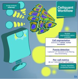
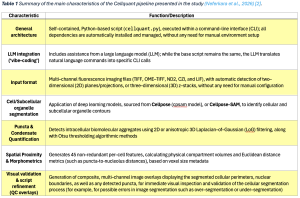
Cellquant is a single Python–based script, assembled with the intention of multi–channel, fluorescence image analysis. The pipeline includes iterative assistance from a large language model (LLM), enabling scientists with limited computer science or programming expertise to perform reproducible, automated, computational imaging analyses (Neferkara et al., 2026) [2]. The pipeline carries out:
- Cell segmentation
- Subcellular localization/Co–localization analysis
- Morphometric quantification
- Spatial proximity calculations across 2 or 3 dimensions
Key aspects of the study
- Stress granule formation was induced in HCT116 cells, via the addition of Arsenite; as Arsenite triggers the phosphorylation of the translation initiation factor eIF2α, blocking protein translation, the stalled ribonucleoproteins condense into G3BP1/2-positive stress granules that recruit additional RNA–binding proteins such as PABPC1. Additional stress granule formation markers such as G3BP1 and G3BP2, which direct their formation in response to increased cytoplasmic concentrations of cytoplasmic mRNA, could also be identified.
- Images of stress granule formation were procured as multi–channel z–stacks, with 3–4 biological replicates per condition; application of Cellquant successfully identified cell and nuclear boundaries, as well as stress granules.
- Budding yeast was cultured across 5 different temperature systems, from 25º to 40ºC; these cells were induced to co–express three proteins, each one tagged with a specific fluorescent marker. These proteins included:
- Tif6, a 60S ribosome biogenesis protein, tagged with a HaloTag (Tif6–Halo), to which the chemical fluorophore JF646 can attach,
- Nsr1, the yeast nucleolin homolog and nucleolar marker tagged with the red fluorescent protein mScarlet–I,
- Sis1, a J–domain protein Hsp40 co–chaperone that forms condensates during heat shock and associates with the nucleolar periphery, tagged with the yellow fluorescent protein mVenus.
- Images of budding yeast were acquired, with segmentation carried out with the help of Cellpose (2D Cellpose masks were linked across the z–stack); nucleolar segmentation was then successfully carried out, based on the fluorescent signal of Nsr1, capturing cellular changes, the localization of the various proteins, and nucleolar reorganization that occurs in yeast due to the alterations in temperature (Neferkara et al., 2026) [2] (Table 1).
Why this work is interesting
The work by Neferkara et al., 2026, presents a valuable and useful toolkit, through which open–source software, tools (Cellpose (cpsam model), Cellpose–SAM) and other packages (scikit–image, numpy, opencv–python–headless, pandas, matplotlib, scipy, PyYAML, pytorch) have been assembled into one Python–based tool, executed from the command–line interface (CLI). The tool also includes iterative LLM assistance, allowing for the generation of reproducible, cellular image analyses, even for scientists without the appropriate familiarity with similar tools. Most importantly, this is all achieved through a standardized, computational format, ‘standardized vibe–coding’, as the authors call it (Neferkara et al., 2026) [2].
Questions to the authors:
- Regarding the implementation of ‘standardized code/LLM input’ paradigm:
- LLM–based agents are frequently updated; how can the user prevent the generation of inconsistent analysis results, or parameters for the exact same biological image analysis query, over time, other than including associated parameters for each run in the provenance.json sidecar?
- Furthermore, are there any LLM agents that you would suggest using with this Python–based script for optimal results, even though all parameters of the run are recorded automatically?
References
[1] Pachitariu M, Stringer C. Cellpose 2.0: how to train your own model. Nat Methods. 2022 Dec;19(12):1634-1641. doi: 10.1038/s41592-022-01663-4
[2] Neferkara A, Winner LA, Ali A, Pincus D. Cellquant: a vibecoder’s guide to image analysis. bioRxiv. 2026 Sep; 710634. doi: https://doi.org/10.64898/2026.03.09.710634
Read preprint