


Explosive cytotoxicity of ‘ruptoblasts’ bridges hormonal surveillance and immune defense
Posted on: 27 October 2025 , updated on: 29 October 2025
Preprint posted on 17 September 2025
Newly discovered cytotoxic cell type in planarians carry out explosive, cross-species killing.
Selected by Inês CaiadoCategories: immunology
Why this preprint matters?
Cytotoxicity is a defence mechanism against cancer and infectious diseases. In addition to directly reducing target cell burden, it promotes antigen release and therefore facilitates the initiation of adaptive immune responses. Despite its importance, most research focuses on immune-mediated cytotoxicity in mammals, overlooking alternative mechanisms in basal animals that could inform immune cell engineering and enhance immunotherapies.
This study identifies a novel cytotoxic cell type in planarian flatworms that induces cell death through a previously undescribed mechanism. The authors use diverse experimental approaches to characterize this cell population and demonstrate its cross-species activity. This preprint highlights how research on basal animals can both advance therapeutic strategies while providing insights to the evolution of immune functions in multicellular organisms.
Background and Hypothesis
Cytotoxicity is critical for the elimination of infected, aberrant, or malignant cells. In animals, this function is primarily mediated by immune effector cells, such as cytotoxic T lymphocytes. In mammals, cytotoxic T cells recognize autoantigens generated during hormone secretion and eliminate hypersecreting cells. This function is essential for maintaining immune surveillance of highly proliferative endocrine tissues, where frequent cell division increases mutation risk. Basal animals, such as planarian flatworms, face similar risks due to their regenerative tissues enriched with proliferative somatic stem cells, known as neoblasts. However, these animals do not possess an adaptive immune system, posing the question: How are aberrant hormone-secreting cells eliminated in planarian flatworms?
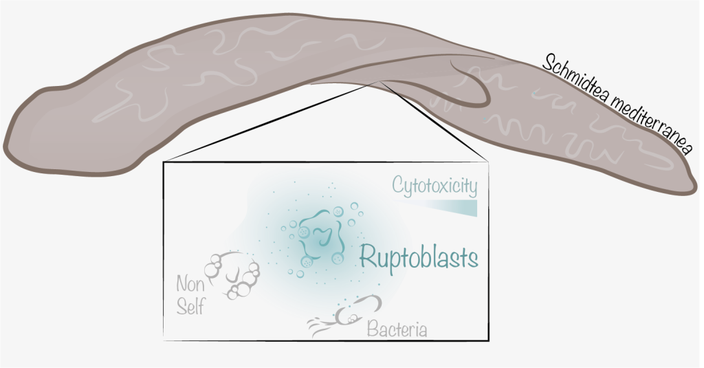
In this preprint, the authors propose that killing of mutated neoblasts is mediated by an unrecognized cytotoxic cell lineage. To test this, they investigated the highly regenerative planarian flatworm Schmidtea mediterranea and focused on the hormone activin, whose levels should be stably maintained to enable regeneration, reproduction, and tissue homeostasis.
Main Findings
- Activin acts as an inflammatory cytokine in planarians. Injection of recombinant activin (ACT-2) activated Smad2/3 and p38 signalling, causing local tissue lesions and cell death. These effects were suppressed if activin receptors or p38 were knocked down. Similarly, genetic chimeras between asexual and sexual planarians developed chronic inflammation with elevated p-Smad2/3 and p-p38 protein levels, suggesting that activin overactivation drives this response.
- Ruptoblasts lyse explosively upon ACT-2 exposure and kill nearby cells. A subset of planarian cells, which the authors named ruptoblasts, lysed explosively within ~2 minutes of ACT-2 exposure in ex vivo conditions. Lysis was dose-dependent and did not occur when cells were treated with control proteins. After lysis, all cells near the ruptoblast were killed (~100 μm), exceeding the granule dispersal range (~30 μm) and therefore confirming that soluble cytotoxic agents are released.
- Ruptoblasts are a glandular/secretory cell type. Ruptoblasts expressed known markers of parenchymal glandular / secretory cells and the transcription factor (TF) fer3l-1 but not act-2 on Single Cell RNA-seq data. Importantly, cathepsin+ phagocytic cells were not responsible for cytotoxicity, as shown by knockdown of cell type specific TFs by RNAi.
- Ruptoblasts are widely distributed in the planarian body and have broad cytotoxicity in vivo. Ruptoblasts were distributed body-wide, except in the head. After ACT-2-induced death, lost ruptoblasts were rapidly regenerated from neoblasts. Deletion of ruptoblasts, by fer3l-1 TF knockdown, suppressed chronic inflammation in chimeras, confirming the central role of ruptoblast in the inflammatory response and tissue damage induced by ACT-2.
- Ruptoblast depletion increases bacterial burden and susceptibility. Depletion of ruptoblasts by act-2 or fer3l-1 RNAi resulted in lower survival post-infection. Incubation of ruptoblasts with GFP-expressing coli only resulted in bacterial death after addition of ACT-2, suggesting that ruptoblasts do not sense bacteria.
- Ruptoblasts kill mammalian cells. After ACT-2 stimulation, lysis of ruptoblasts co-cultured with human embryonic kidney (HEK293) cells or mouse macrophages (RAW264.7) resulted in mammalian cell death within ~200 μm. Similarly, the supernatant from activin-induced ruptosis was cytotoxic, whereas mechanically lysed ruptoblasts without ACT-2 activation were not, indicating activin-dependent activation of the killing agents.
- Ruptoblasts lysis (Ruptosis) is regulated by intracellular calcium and actomyosin cytoskeletal dynamics. Chelating intracellular calcium prevented full membrane rupture but left the actin cytoskeleton intact, while nuclear disruption proceeded as in controls. This suggests calcium is dispensable for initiating ruptosis but essential for complete cytoskeletal disassembly and membrane disintegration. Inhibition of myosin II or actin polymerization delayed ruptosis and prevented nucleus rupture. Conversely, stabilizing actin accelerated nucleus dissociation, indicating that actin is essential for maintaining the pressure and releasing it at the proper point to achieve explosiveness.

Figure 1- Mechanism of Ruptosis. Ruptoblasts are activated by the hormone activin, inducing rapid and explosive cell lysis. This process is characterized by synchronized membrane rupture and actin depolymerization, leading to the release of cytotoxic granules.
Key conclusions
In short, this preprint reports the discovery of a previously unrecognized cytotoxic cell type in planarians that is activated by the hormone activin and kills through a new cell death form. These cells act with remarkable speed and show cross-species reactivity. Their mechanism of action is initiated by actin, triggering intracellular free calcium signalling and driving energy buildup that is likely released upon cytoskeletal disassembly, causing the explosive lysis phenotype.
Questions
- In the killing assays for planarian cells, you report a killing range of ~100 μm, while for mammalian cells you mention ~200 μm. Is this consistent? Do you expect mammalian cells to be more sensitive to ruptosis?
- You hypothesized that phagocytic cells could be activin-producers. However, in bacterial killing assays, P5 cell cultures-which based on scRNA-seq include both phagocytic cells and ruptoblasts- required exogenous ACT-2 to induce bacterial death. Do you attribute this to insufficient phagocyte numbers or to the assay duration being too short to accumulate high levels of activin? Which other cell types could produce activin?
doi: https://doi.org/10.1242/prelights.41834
Read preprint (No Ratings Yet)
Sign up to customise the site to your preferences and to receive alerts
Register hereAlso in the immunology category:
Circadian Clock Programming of Anticipatory Antiviral Immunity Gates Enteric Virus Infection Susceptibility
Owen Ang
Inhibition of VP2-mediated entry: a potential antiviral strategy to treat or prevent calicivirus disease
Orestis Savva
EBV reprograms autoreactive anti-CNS B cells as antigen presenting cells in multiple sclerosis
Léa Bastien et al.
preLists in the immunology category:
BSDB Spring Meeting: Molecules to Morphogenesis
The British Society for Developmental Biology (BSDB) Spring Meeting Molecules to Morphogenesis was held from 23–26 March 2026 at the University of Warwick (UK). This meeting brought together a vibrant community of researchers to discuss how molecular mechanisms are integrated across scales to drive morphogenesis, spanning diverse model systems and approaches. This preList contains preprints by presenters from the talk and poster sessions at the meeting. Please do get in touch at preLights@biologists.com if you notice any relevant preprints that we may have missed.
| List by | Ingrid Tsang |
SciELO preprints – From 2025 onwards
SciELO has become a cornerstone of open, multilingual scholarly communication across Latin America. Its preprint server, SciELO preprints, is expanding the global reach of preprinted research from the region (for more information, see our interview with Carolina Tanigushi). This preList brings together biological, English language SciELO preprints to help readers discover emerging work from the Global South. By highlighting these preprints in one place, we aim to support visibility, encourage early feedback, and showcase the vibrant research communities contributing to SciELO’s open science ecosystem.
| List by | Carolina Tanigushi |
Community-driven preList – Immunology
In this community-driven preList, a group of preLighters, with expertise in different areas of immunology have worked together to create this preprint reading list.
| List by | Felipe Del Valle Batalla et al. |
Journal of Cell Science meeting ‘Imaging Cell Dynamics’
This preList highlights the preprints discussed at the JCS meeting 'Imaging Cell Dynamics'. The meeting was held from 14 - 17 May 2023 in Lisbon, Portugal and was organised by Erika Holzbaur, Jennifer Lippincott-Schwartz, Rob Parton and Michael Way.
| List by | Helen Zenner |
Fibroblasts
The advances in fibroblast biology preList explores the recent discoveries and preprints of the fibroblast world. Get ready to immerse yourself with this list created for fibroblasts aficionados and lovers, and beyond. Here, my goal is to include preprints of fibroblast biology, heterogeneity, fate, extracellular matrix, behavior, topography, single-cell atlases, spatial transcriptomics, and their matrix!
| List by | Osvaldo Contreras |
Single Cell Biology 2020
A list of preprints mentioned at the Wellcome Genome Campus Single Cell Biology 2020 meeting.
| List by | Alex Eve |
Autophagy
Preprints on autophagy and lysosomal degradation and its role in neurodegeneration and disease. Includes molecular mechanisms, upstream signalling and regulation as well as studies on pharmaceutical interventions to upregulate the process.
| List by | Sandra Malmgren Hill |
Antimicrobials: Discovery, clinical use, and development of resistance
Preprints that describe the discovery of new antimicrobials and any improvements made regarding their clinical use. Includes preprints that detail the factors affecting antimicrobial selection and the development of antimicrobial resistance.
| List by | Zhang-He Goh |
Zebrafish immunology
A compilation of cutting-edge research that uses the zebrafish as a model system to elucidate novel immunological mechanisms in health and disease.
| List by | Shikha Nayar |