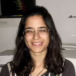
My name is Mariana De Niz. I am a postdoctoral fellow at the Institute of Molecular Medicine in Lisbon, Portugal. My work focuses on understanding the biophysics and dynamics of the parasite Trypanosoma brucei (causative of sleeping sickness), in various tissues of mammalian hosts. I am particularly interested in microscopy methods to image host-parasite interactions, and am keen on developing new methods that enable visualising and investigating these interactions. In my PhD and first postdoc, I worked on investigating various stages of the malaria-causative parasite Plasmodium. I was particularly interested in understanding parasite tropism and mechanisms of sequestration during Plasmodium blood stages, and host cell organelle manipulation during Plasmodium liver stage development.
Creating Clear and Informative Image-based Figures for Scientific Publications
Mariana De Niz
Blocking palmitoylation of Toxoplasma gondii myosin light chain 1 disrupts glideosome composition but has little impact on parasite motility
Mariana De Niz
A mating-induced reproductive gene promotes Anopheles tolerance to Plasmodium falciparum infection
Mariana De Niz
A zebrafish model for COVID-19 recapitulates olfactory and cardiovascular pathophysiologies caused by SARS-CoV-2
Mariana De Niz
Pocket MUSE: an affordable, versatile and high performance fluorescence microscope using a smartphone
Mariana De Niz
Nuclear stiffness decreases with disruption of the extracellular matrix in living tissues
Mariana De Niz
Imaging transmembrane dynamics of biomolecules at live cell plasma membranes using quenchers in extracellular environment
Mariana De Niz
“Viscotaxis”- Directed Migration of Mesenchymal Stem Cells in Response to Loss Modulus Gradient
Mariana De Niz
FLIMJ: an open-source ImageJ toolkit for fluorescence lifetime image data analysis
Mariana De Niz
Deep learning-enhanced light-field imaging with continuous validation
Mariana De Niz
Interstitial spaces are continuous across tissue and organ boundaries in humans
Mariana De Niz
3D quantification of zebrafish cerebrovascular architecture by automated image analysis of light sheet fluorescence microscopy datasets
Mariana De Niz


