Spatial heterogeneity of cell-matrix adhesive forces predicts human glioblastoma migration
Posted on: 17 June 2020
Preprint posted on 8 May 2020
Article now published in Neuro-Oncology Advances at http://dx.doi.org/10.1093/noajnl/vdaa081
Categories: biophysics, cancer biology
Background
Glioblastoma (GBM) is a highly aggressive and currently incurable brain tumor. The main cause of mortality in GBM patients is the invasive rim of cells migrating away from the main tumor mass, and invading healthy parts of the brain. This extensive and infiltrative growth pattern makes surgical resection difficult, and limits the efficacy of radiation therapy.
While it is known that cell migration and invasion are driven by mechanical forces, our current understanding of the physical factors involved in glioma infiltration remains limited. It is known that GBM display a mesenchymal mode of migration, using focal adhesion proteins as molecular clutches to transmit force to their environments. Although attractive, so far, therapeutics designed to target adhesion receptors or proteases have failed in clinical trials. It is thought that this failure might be due to the heterogeneity in expression of the adhesion proteins both, between patients, and between tumour regions in the same patient. In their work, Rezk and colleagues for the first time investigated adhesive and migratory properties of different GBM subpopulations from different patients, and different regions within the tumours.

Key findings and developments
In this study, Rezk and colleagues investigated the adhesion properties within and between patients’ tumors on a cellular level and tested whether these properties correlate with cell migration. For this purpose, the authors used 5-aminolevulinic acid (5-ALA) fluorescence guided therapy in various patients to separate different tumour sections for further study. These sections included the tumour core (highest fluorescence intensity), the tumour rim (medium fluorescence intensity), and tumour margins (not fluorescent). The authors then adapted a microfluidic device to detach adherent cells through shear stress, and cultured them in PDMS with a low modulus consistent with the stiffness of the environment that GBM infiltrate.
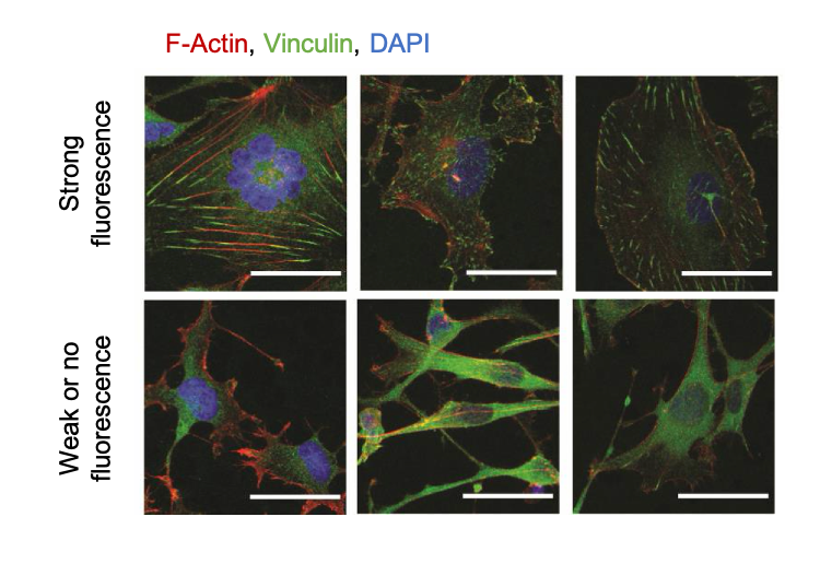
Cell spreading area is known to be an indicator of how cells mechanically interact with their extracellular environment. The authors detected morphological differences in cells derived from different patients, and demonstrated that morphological heterogeneity within and between tumours was related to 5-ALA fluorescence intensity (i.e. tumour region). To investigate whether cell morphology relates to the way cells adhere to their environment, the authors went on to explore specific patterns of key cytoskeletal proteins. For this, they imaged the localization and structure of actin filaments and the actin-binding protein vinculin in cells from different tumour regions, and demonstrated that shape and distribution of actin and vinculin differed between cells derived from each region.
The authors propose that the organization of actin filaments and vinculin suggests spatial differences in focal adhesion assembly and enlargement between cells of different tumour regions. The authors went on to build a microfluidic device to quantitate cell matrix adhesion strength. Cells were injected into channels and subjected to a controlled flow rate to create a constant shear force on the cell. The output of this setup is quantification of the fraction of detached cells over time. For the purpose of the work, the authors took measurements of cell detachment after 5 minutes of flow, for quantifications of adhesion strength. They found that cells derived from areas corresponding to the tumour rim and margins had significantly lower cell matrix adhesion and smaller spreading areas, than cells derived from the tumour core.
They then quantified the migratory behaviour of cells derived from the different tumour regions. For this, the trajectory of the different cell populations on compliant PDMS substrate was recorded. This showed differences in migratory capacity between cells derived from different patients, and importantly, it showed that cells derived from the tumour core were significantly slower than cells derived from the tumour rim or margins.
Altogether, the authors conclude that cell-adhesion strength could be an accurate predictor of tumour cell migration, unlike molecular classification which have so far failed to provide accurate predictions. They further suggest that preclinical tests aimed at developing anti-invasive drugs or adhesion inhibitors against GBM, would be more accurate if using cell lines derived from the tumour periphery, rather than the tumour core.
What I like about this preprint
I like this preprint because it investigates a new aspect of GBM, and aims to determine the source of the heterogeneity that so far has hindered treatment, and the focus of clinical trials. I like inter-disciplinary research and I like approaches that are out-of-the-box to address scientific questions. I think this work beautifully does this. Also, I think there are pieces of work in multiple fields showing more and more the importance of biophysics and biomechanics. For me this is a new era of research, in which we no longer only focus on the molecular and OMICS, but have broadened our approach and questions.
References
- Rezk R, et al, Spatial heterogeneity of cell-matrix adhesive forces predicts human glioblastoma migration, bioRxiv, 2020
- Heinrich MA, et al, Tissue size controls patterns of cell proliferation and migration in freely expanding epithelia, bioRxiv, 2020.
- Plodinec, Marija, et al. The nanomechanical signature of breast cancer, Nature nanotechnology, 11, 2012.
doi: https://doi.org/10.1242/prelights.21979
Read preprintSign up to customise the site to your preferences and to receive alerts
Register hereAlso in the biophysics category:
Active flows drive clustering and sorting of membrane components with differential affinity to dynamic actin cytoskeleton
Teodora Piskova
Junctional Heterogeneity Shapes Epithelial Morphospace
Bhaval Parmar
Mitochondrial Fatty Acid Oxidation is Stimulated by Red Light Irradiation
Rickson Ribeiro, Marcus Oliveira
Also in the cancer biology category:
A Novel Chimeric Antigen Receptor (CAR) - Strategy to Target EGFRVIII-Mutated Glioblastoma Cells via Macrophages
Dina Kabbara
Taxane-Induced Conformational Changes in the Microtubule Lattice Activate GEF-H1-Dependent RhoA Signaling
Vibha SINGH
ROCK2 inhibition has a dual role in reducing ECM remodelling and cell growth, while impairing migration and invasion
Sharvari Pitke
preLists in the biophysics category:
October in preprints – DevBio & Stem cell biology
Each month, preLighters with expertise across developmental and stem cell biology nominate a few recent developmental and stem cell biology (and related) preprints they’re excited about and explain in a single paragraph why. Short, snappy picks from working scientists — a quick way to spot fresh ideas, bold methods and papers worth reading in full. These preprints can all be found in the October preprint list published on the Node.
| List by | Deevitha Balasubramanian et al. |
October in preprints – Cell biology edition
Different preLighters, with expertise across cell biology, have worked together to create this preprint reading list for researchers with an interest in cell biology. This month, most picks fall under (1) Cell organelles and organisation, followed by (2) Mechanosignaling and mechanotransduction, (3) Cell cycle and division and (4) Cell migration
| List by | Matthew Davies et al. |
March in preprints – the CellBio edition
A group of preLighters, with expertise in different areas of cell biology, have worked together to create this preprint reading lists for researchers with an interest in cell biology. This month, categories include: 1) cancer biology 2) cell migration 3) cell organelles and organisation 4) cell signalling and mechanosensing 5) genetics and genomics 6) other
| List by | Girish Kale et al. |
Biologists @ 100 conference preList
This preList aims to capture all preprints being discussed at the Biologists @100 conference in Liverpool, UK, either as part of the poster sessions or the (flash/short/full-length) talks.
| List by | Reinier Prosee, Jonathan Townson |
February in preprints – the CellBio edition
A group of preLighters, with expertise in different areas of cell biology, have worked together to create this preprint reading lists for researchers with an interest in cell biology. This month, categories include: 1) biochemistry and cell metabolism 2) cell organelles and organisation 3) cell signalling, migration and mechanosensing
| List by | Barbora Knotkova et al. |
preLights peer support – preprints of interest
This is a preprint repository to organise the preprints and preLights covered through the 'preLights peer support' initiative.
| List by | preLights peer support |
66th Biophysical Society Annual Meeting, 2022
Preprints presented at the 66th BPS Annual Meeting, Feb 19 - 23, 2022 (The below list is not exhaustive and the preprints are listed in no particular order.)
| List by | Soni Mohapatra |
EMBL Synthetic Morphogenesis: From Gene Circuits to Tissue Architecture (2021)
A list of preprints mentioned at the #EESmorphoG virtual meeting in 2021.
| List by | Alex Eve |
Biophysical Society Meeting 2020
Some preprints presented at the Biophysical Society Meeting 2020 in San Diego, USA.
| List by | Tessa Sinnige |
ASCB EMBO Annual Meeting 2019
A collection of preprints presented at the 2019 ASCB EMBO Meeting in Washington, DC (December 7-11)
| List by | Madhuja Samaddar et al. |
EMBL Seeing is Believing – Imaging the Molecular Processes of Life
Preprints discussed at the 2019 edition of Seeing is Believing, at EMBL Heidelberg from the 9th-12th October 2019
| List by | Dey Lab |
Biomolecular NMR
Preprints related to the application and development of biomolecular NMR spectroscopy
| List by | Reid Alderson |
Biophysical Society Annual Meeting 2019
Few of the preprints that were discussed in the recent BPS annual meeting at Baltimore, USA
| List by | Joseph Jose Thottacherry |
Also in the cancer biology category:
October in preprints – Cell biology edition
Different preLighters, with expertise across cell biology, have worked together to create this preprint reading list for researchers with an interest in cell biology. This month, most picks fall under (1) Cell organelles and organisation, followed by (2) Mechanosignaling and mechanotransduction, (3) Cell cycle and division and (4) Cell migration
| List by | Matthew Davies et al. |
September in preprints – Cell biology edition
A group of preLighters, with expertise in different areas of cell biology, have worked together to create this preprint reading list. This month, categories include: (1) Cell organelles and organisation, (2) Cell signalling and mechanosensing, (3) Cell metabolism, (4) Cell cycle and division, (5) Cell migration
| List by | Sristilekha Nath et al. |
July in preprints – the CellBio edition
A group of preLighters, with expertise in different areas of cell biology, have worked together to create this preprint reading lists for researchers with an interest in cell biology. This month, categories include: (1) Cell Signalling and Mechanosensing (2) Cell Cycle and Division (3) Cell Migration and Cytoskeleton (4) Cancer Biology (5) Cell Organelles and Organisation
| List by | Girish Kale et al. |
June in preprints – the CellBio edition
A group of preLighters, with expertise in different areas of cell biology, have worked together to create this preprint reading lists for researchers with an interest in cell biology. This month, categories include: (1) Cell organelles and organisation (2) Cell signaling and mechanosensation (3) Genetics/gene expression (4) Biochemistry (5) Cytoskeleton
| List by | Barbora Knotkova et al. |
May in preprints – the CellBio edition
A group of preLighters, with expertise in different areas of cell biology, have worked together to create this preprint reading lists for researchers with an interest in cell biology. This month, categories include: 1) Biochemistry/metabolism 2) Cancer cell Biology 3) Cell adhesion, migration and cytoskeleton 4) Cell organelles and organisation 5) Cell signalling and 6) Genetics
| List by | Barbora Knotkova et al. |
Keystone Symposium – Metabolic and Nutritional Control of Development and Cell Fate
This preList contains preprints discussed during the Metabolic and Nutritional Control of Development and Cell Fate Keystone Symposia. This conference was organized by Lydia Finley and Ralph J. DeBerardinis and held in the Wylie Center and Tupper Manor at Endicott College, Beverly, MA, United States from May 7th to 9th 2025. This meeting marked the first in-person gathering of leading researchers exploring how metabolism influences development, including processes like cell fate, tissue patterning, and organ function, through nutrient availability and metabolic regulation. By integrating modern metabolic tools with genetic and epidemiological insights across model organisms, this event highlighted key mechanisms and identified open questions to advance the emerging field of developmental metabolism.
| List by | Virginia Savy, Martin Estermann |
April in preprints – the CellBio edition
A group of preLighters, with expertise in different areas of cell biology, have worked together to create this preprint reading lists for researchers with an interest in cell biology. This month, categories include: 1) biochemistry/metabolism 2) cell cycle and division 3) cell organelles and organisation 4) cell signalling and mechanosensing 5) (epi)genetics
| List by | Vibha SINGH et al. |
March in preprints – the CellBio edition
A group of preLighters, with expertise in different areas of cell biology, have worked together to create this preprint reading lists for researchers with an interest in cell biology. This month, categories include: 1) cancer biology 2) cell migration 3) cell organelles and organisation 4) cell signalling and mechanosensing 5) genetics and genomics 6) other
| List by | Girish Kale et al. |
Biologists @ 100 conference preList
This preList aims to capture all preprints being discussed at the Biologists @100 conference in Liverpool, UK, either as part of the poster sessions or the (flash/short/full-length) talks.
| List by | Reinier Prosee, Jonathan Townson |
February in preprints – the CellBio edition
A group of preLighters, with expertise in different areas of cell biology, have worked together to create this preprint reading lists for researchers with an interest in cell biology. This month, categories include: 1) biochemistry and cell metabolism 2) cell organelles and organisation 3) cell signalling, migration and mechanosensing
| List by | Barbora Knotkova et al. |
BSCB-Biochemical Society 2024 Cell Migration meeting
This preList features preprints that were discussed and presented during the BSCB-Biochemical Society 2024 Cell Migration meeting in Birmingham, UK in April 2024. Kindly put together by Sara Morais da Silva, Reviews Editor at Journal of Cell Science.
| List by | Reinier Prosee |
CSHL 87th Symposium: Stem Cells
Preprints mentioned by speakers at the #CSHLsymp23
| List by | Alex Eve |
Journal of Cell Science meeting ‘Imaging Cell Dynamics’
This preList highlights the preprints discussed at the JCS meeting 'Imaging Cell Dynamics'. The meeting was held from 14 - 17 May 2023 in Lisbon, Portugal and was organised by Erika Holzbaur, Jennifer Lippincott-Schwartz, Rob Parton and Michael Way.
| List by | Helen Zenner |
CellBio 2022 – An ASCB/EMBO Meeting
This preLists features preprints that were discussed and presented during the CellBio 2022 meeting in Washington, DC in December 2022.
| List by | Nadja Hümpfer et al. |
Fibroblasts
The advances in fibroblast biology preList explores the recent discoveries and preprints of the fibroblast world. Get ready to immerse yourself with this list created for fibroblasts aficionados and lovers, and beyond. Here, my goal is to include preprints of fibroblast biology, heterogeneity, fate, extracellular matrix, behavior, topography, single-cell atlases, spatial transcriptomics, and their matrix!
| List by | Osvaldo Contreras |
Single Cell Biology 2020
A list of preprints mentioned at the Wellcome Genome Campus Single Cell Biology 2020 meeting.
| List by | Alex Eve |
ASCB EMBO Annual Meeting 2019
A collection of preprints presented at the 2019 ASCB EMBO Meeting in Washington, DC (December 7-11)
| List by | Madhuja Samaddar et al. |
Lung Disease and Regeneration
This preprint list compiles highlights from the field of lung biology.
| List by | Rob Hynds |
Anticancer agents: Discovery and clinical use
Preprints that describe the discovery of anticancer agents and their clinical use. Includes both small molecules and macromolecules like biologics.
| List by | Zhang-He Goh |


 (No Ratings Yet)