


Synapses drive local mitochondrial ATP synthesis to fuel plasticity
Posted on: 2 May 2025
Preprint posted on 10 April 2025
New spine-mitochondrial ATP ratiometric reporter shows that synaptic activity triggers a rapid, sustained ATP boost in dendritic spines, fueled by local mitochondria.
Selected by Felipe Del Valle BatallaCategories: neuroscience
Background: Our brains are constantly forming and stabilizing memories, a process that relies on synaptic plasticity. Supplying ATP to these synapses, which can be located hundreds (or even thousands) of microns away from the neuronal cell body, poses a significant challenge. Therefore, synapses require an immediate and local energy supply, typically provided by mitochondria stabilized near dendritic spines. While it is known that mitochondrial dysfunction is associated with various neurological disorders, the fundamental mechanisms underlying dendritic spine energetics and how synapses signal their energy needs to local mitochondria have remained largely unknown. The primary obstacle in understanding spine energetics has been the lack of sensitive tools to measure ATP within small compartments like spines and mitochondria, coupled with the need to combine these measurements with single-spine stimulation and plasticity induction.
This preprint addresses these challenges by employing newly engineered spine- and mitochondria-targeted ATP reporters, along with two-photon glutamate uncaging, to study ATP dynamics upon synaptic plasticity induction at high resolution.
Key Findings of the preprint:
Spn-ATP: A quantitative reporter of spine ATP
The researchers begin with the introduction of a new quantitative spine ATP reporter called Spn-ATP. This reporter consists of luciferase (luc) fused to the postsynaptic protein Homer2 and the pH-sensitive fluorescent protein mOrange2 (mOrg) for ratiometric measurements (Fig.2A). Due to the low light emission of luminescence reporters, the authors custom-built an inverted spinning disk confocal microscope with specialized cameras for sensitive luminescence imaging and simultaneous two-photon manipulation. Using this setup and a calibrated Spn-ATP reporter, they measured steady-state ATPspine concentrations at 0.41 ± 0.01 mM in areas 50–100 µm from the soma. They also found that steady-state ATPspine decreases with increasing distance from the soma (Fig.2B).

Fig. 2 from the preprint: Synaptic plasticity induction increases ATPspine.
(A) Illustration showing Spn-ATP for ATPspine measurements following synaptic plasticity induction (lightning bolt). (B) Representative images showing spine calcium (white, GCaMP6s) and Spn-ATP luminescence (orange, luc) increase upon synaptic plasticity induction (white and black asterisk). The intensity scale is in arbitrary units. Image made available under a CC-BY-NC-ND 4.0 International license.
Synaptic plasticity induction increases ATPspine and drives local ATPmito synthesis
Contrary to expectations of ATP depletion, synaptic plasticity induction via two-photon glutamate uncaging at individual spines led to an instant (~1 min) and sustained (~11+ min) increase in ATPspine of about 1.5-fold. This increase was primarily fueled by mitochondrial oxidative phosphorylation initially, while both oxidative phosphorylation and glycolysis contributed to the sustained increase. Inhibition of these pathways affected the ATPspine increase and impaired spine structural plasticity. This instant and sustained energy supply is considered essential for synaptic plasticity. The increase in ATPspine was spatially restricted to spines within 10 µm of the stimulated spine, pointing to a local ATP source, likely mitochondria.
To investigate the mitochondrial role, the authors developed Mito-ATP (Fig.3A), a reporter targeting luciferase to the mitochondrial matrix for ratiometric ATP measurements. Using Mito-ATP, they observed a rapid rise in ATPmito within the first two minutes of synaptic plasticity induction, which was sustained for at least 12 minutes. This increase was dependent on mitochondrial oxidative phosphorylation and, importantly, was spatially restricted to a ~10 µm compartment within mitochondria near the stimulated spine.
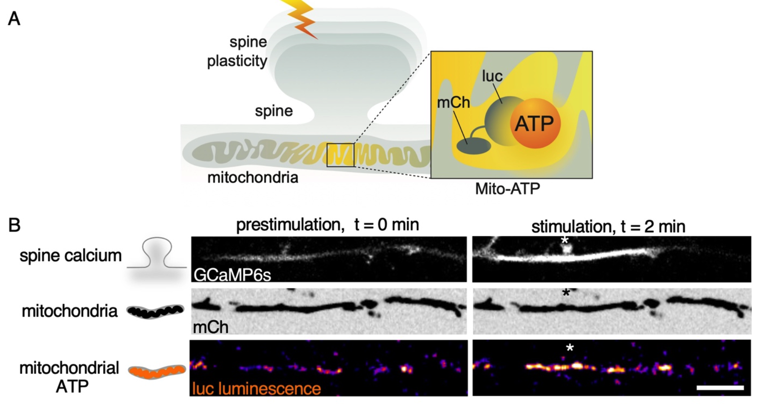
Fig. 3 from the preprint. (A) Illustration showing the design of the mitochondrial ATP sensor, Mito-ATP, for ATPmito measurements following synaptic plasticity induction (lightning bolt). Luciferase (luc) is conjugated to the fluorescent protein mCherry2 (mCh) and targeted into the mitochondrial matrix. (B) Representative images showing spine calcium influx in the plasticity-induced spine (white and black asterisk) measured using GCaMP6s fluorescence. In response to spine plasticity induction, there is a spatially restricted increase in mitochondrial luc luminescence at the base of the plasticity-induced spine without a change in the mCh fluorescence. Scale bar 10 µm. Image made available under a CC-BY-NC-ND 4.0 International license.
ER plays a regulatory, but not obligatory, role in mitochondrial calcium influx
Having established a method for quantifying steady-state ATP levels at the spine and observing their spatial gradient, the researchers next sought to investigate how synaptic activity and plasticity events dynamically alter these local ATP concentrations. Synaptic plasticity induction increases local mitochondrial calcium influx. This study explored the link between synaptic inputs and mitochondrial ATP synthesis, hypothesizing that calcium, known to activate Krebs cycle enzymes, could be the signal.
To test the hypothesis this, they used special tools to track calcium levels in both the cytoplasm and the mitochondria of neurons. When they stimulated a single synapse with via glutamate uncaging they observed a rapid and simultaneous increase in calcium within the nearby mitochondria.
This mitochondrial calcium influx had specific requirements: it depended on the activation of NMDAR receptors at the synapse and only occurred if the calcium concentration in the dendrite, right at the base of the stimulated spine, reached a certain threshold. Interestingly, this calcium uptake by mitochondria was very localized, happening only within a small region (around 20 micrometers) of the stimulated synapse. This result suggests that calcium acts as a fast, local, and sustained signal that tells the mitochondria to produce more ATP specifically for that active spine (Fig.7).
The authors also looked at the role of the endoplasmic reticulum (ER), another cellular compartment that stores calcium. By interfering with the ER’s ability to take up or release calcium, they found only a minor impact on the mitochondrial calcium uptake following synaptic stimulation. This observation indicates that while the ER might have some influence, it’s not essential for this process of mitochondrial calcium entry triggered by synaptic activity. This was further confirmed by the fact that their standard stimulation method didn’t effectively release calcium from the ER, and because many spines don’t have much ER present within them.

Fig. 7 from the preprint. Illustration showing the significance of synaptic calcium signal input (green sphere) driving local mitochondrial calcium signaling (green sphere, green cristae) enabled by local mitochondrial stabilization (VAP, actin tethers), resulting in spatially restricted mitochondrial ATP production (yellow cristae) and its distribution to plasticity-induced spines (yellow spines) measured using mitochondrial- (Mito-ATP) and spine-targeted (Spn-ATP) ATP reporters, respectively. Image made available under a CC-BY-NC-ND 4.0 International license.
Why I think this work is important: This preprint presents a significant advance in our understanding of the energetic groundings of synaptic plasticity by allowing for a direct and quantitative measurement of ATP dynamics during synaptic activity and plasticity. Furthermore, the work highlights the critical role of mitochondrial stability in ensuring a reliable local energy supply for sustained synaptic function, with clear implications for neurological disorders. The comparison of steady-state ATP levels and energy dynamics between dendritic spines and presynaptic terminals provides valuable insights into the distinct energetic requirements of these neuronal compartments.
doi: https://doi.org/10.1242/prelights.40383
Read preprint (No Ratings Yet)
Sign up to customise the site to your preferences and to receive alerts
Register hereAlso in the neuroscience category:
Behavioral characteristics of an extremely old rhesus macaque in a zoo: Dementia-like symptoms and implications for quality of life of geriatric animals
Stefan Friedrich Wirth
EBV reprograms autoreactive anti-CNS B cells as antigen presenting cells in multiple sclerosis
Léa Bastien et al.
The Endocannabinoid System’s Contribution to Placebo Analgesia
Thomas Nicodemo Arrieta et al.
preLists in the neuroscience category:
preLighters’ choice – Handpicked DevBio preprints
preLighters with expertise across developmental and stem cell biology have nominated a few developmental biology (and related) preprints they’re excited about and explain in a few paragraph why. Concise preprint highlights, prepared by the preLighter community – a quick way to spot upcoming trends, new methods and fresh ideas.
| List by | Theodora Stougiannou et al. |
BSDB Spring Meeting: Molecules to Morphogenesis
The British Society for Developmental Biology (BSDB) Spring Meeting Molecules to Morphogenesis was held from 23–26 March 2026 at the University of Warwick (UK). This meeting brought together a vibrant community of researchers to discuss how molecular mechanisms are integrated across scales to drive morphogenesis, spanning diverse model systems and approaches. This preList contains preprints by presenters from the talk and poster sessions at the meeting. Please do get in touch at preLights@biologists.com if you notice any relevant preprints that we may have missed.
| List by | Ingrid Tsang |
Keystone Symposium on Stem Cell Models in Embryology 2026
The Keystone Symposium on Stem Cell Models in Embryology, 2026, was organised by Jun Wu (UT Southwestern), Jianping Fu (University of Michigan) and Miki Ebisuya (TU Dresden) and held at Asilomar Conference Grounds in California (US). The meeting discussed recent advances made in establishing stem-cell-based embryo models, what fundamental insights into developmental processes have been gleaned from them, as well as how they are beginning to be applied more widely. This prelist contains preprints by presenters at the talk and poster sessions at the conference, which our Reviews Editor in attendance spotted. Please do reach out to preLights@biologists.com if you notice any that we’ve missed.
| List by | Ingrid Tsang |
November in preprints – DevBio & Stem cell biology
preLighters with expertise across developmental and stem cell biology have nominated a few developmental and stem cell biology (and related) preprints posted in November they’re excited about and explain in a single paragraph why. Concise preprint highlights, prepared by the preLighter community – a quick way to spot upcoming trends, new methods and fresh ideas.
| List by | Aline Grata et al. |
October in preprints – DevBio & Stem cell biology
Each month, preLighters with expertise across developmental and stem cell biology nominate a few recent developmental and stem cell biology (and related) preprints they’re excited about and explain in a single paragraph why. Short, snappy picks from working scientists — a quick way to spot fresh ideas, bold methods and papers worth reading in full. These preprints can all be found in the October preprint list published on the Node.
| List by | Deevitha Balasubramanian et al. |
October in preprints – Cell biology edition
Different preLighters, with expertise across cell biology, have worked together to create this preprint reading list for researchers with an interest in cell biology. This month, most picks fall under (1) Cell organelles and organisation, followed by (2) Mechanosignaling and mechanotransduction, (3) Cell cycle and division and (4) Cell migration
| List by | Matthew Davies et al. |
July in preprints – the CellBio edition
A group of preLighters, with expertise in different areas of cell biology, have worked together to create this preprint reading lists for researchers with an interest in cell biology. This month, categories include: (1) Cell Signalling and Mechanosensing (2) Cell Cycle and Division (3) Cell Migration and Cytoskeleton (4) Cancer Biology (5) Cell Organelles and Organisation
| List by | Girish Kale et al. |
May in preprints – the CellBio edition
A group of preLighters, with expertise in different areas of cell biology, have worked together to create this preprint reading lists for researchers with an interest in cell biology. This month, categories include: 1) Biochemistry/metabolism 2) Cancer cell Biology 3) Cell adhesion, migration and cytoskeleton 4) Cell organelles and organisation 5) Cell signalling and 6) Genetics
| List by | Barbora Knotkova et al. |
April in preprints – the CellBio edition
A group of preLighters, with expertise in different areas of cell biology, have worked together to create this preprint reading lists for researchers with an interest in cell biology. This month, categories include: 1) biochemistry/metabolism 2) cell cycle and division 3) cell organelles and organisation 4) cell signalling and mechanosensing 5) (epi)genetics
| List by | Vibha SINGH et al. |
Biologists @ 100 conference preList
This preList aims to capture all preprints being discussed at the Biologists @100 conference in Liverpool, UK, either as part of the poster sessions or the (flash/short/full-length) talks.
| List by | Reinier Prosee, Jonathan Townson |
2024 Hypothalamus GRC
This 2024 Hypothalamus GRC (Gordon Research Conference) preList offers an overview of cutting-edge research focused on the hypothalamus, a critical brain region involved in regulating homeostasis, behavior, and neuroendocrine functions. The studies included cover a range of topics, including neural circuits, molecular mechanisms, and the role of the hypothalamus in health and disease. This collection highlights some of the latest advances in understanding hypothalamic function, with potential implications for treating disorders such as obesity, stress, and metabolic diseases.
| List by | Nathalie Krauth |
‘In preprints’ from Development 2022-2023
A list of the preprints featured in Development's 'In preprints' articles between 2022-2023
| List by | Alex Eve, Katherine Brown |
CSHL 87th Symposium: Stem Cells
Preprints mentioned by speakers at the #CSHLsymp23
| List by | Alex Eve |
Journal of Cell Science meeting ‘Imaging Cell Dynamics’
This preList highlights the preprints discussed at the JCS meeting 'Imaging Cell Dynamics'. The meeting was held from 14 - 17 May 2023 in Lisbon, Portugal and was organised by Erika Holzbaur, Jennifer Lippincott-Schwartz, Rob Parton and Michael Way.
| List by | Helen Zenner |
FENS 2020
A collection of preprints presented during the virtual meeting of the Federation of European Neuroscience Societies (FENS) in 2020
| List by | Ana Dorrego-Rivas |
ASCB EMBO Annual Meeting 2019
A collection of preprints presented at the 2019 ASCB EMBO Meeting in Washington, DC (December 7-11)
| List by | Madhuja Samaddar et al. |
SDB 78th Annual Meeting 2019
A curation of the preprints presented at the SDB meeting in Boston, July 26-30 2019. The preList will be updated throughout the duration of the meeting.
| List by | Alex Eve |
Autophagy
Preprints on autophagy and lysosomal degradation and its role in neurodegeneration and disease. Includes molecular mechanisms, upstream signalling and regulation as well as studies on pharmaceutical interventions to upregulate the process.
| List by | Sandra Malmgren Hill |
Young Embryologist Network Conference 2019
Preprints presented at the Young Embryologist Network 2019 conference, 13 May, The Francis Crick Institute, London
| List by | Alex Eve |