


Intravital optoacoustic ultrasound bio-microscopy reveals radiation-inhibited skull angiogenesis
Posted on: 13 May 2019 , updated on: 14 May 2019
Preprint posted on 19 February 2019
Article now published in Bone at http://dx.doi.org/10.1016/j.bone.2020.115251
Shedding light on skull and cerebral vascular changes: optoacoustic ultrasound bio-imaging shows irradiation effects on angiogenesis inhibition.
Selected by Mariana De NizCategories: biophysics, cancer biology, pathology, physiology
Background
Optical microscopy has been pivotal for biological discovery for more than three centuries. However, microscopy methods are limited for in vivo tissue imaging by light scattering. Scattering consists on photon absorption and re-emission without loss of energy, but with a change in photon direction which occurs due to photon interaction with cellular structures. Multiple scattering events result in photon diffusion. This ultimately imposes limits to the penetration capacity of microscopic imaging. Additionally, different tissues have different absorption and scattering coefficients, making some tissues more or less amenable to optical imaging techniques (Reviewed in 1).
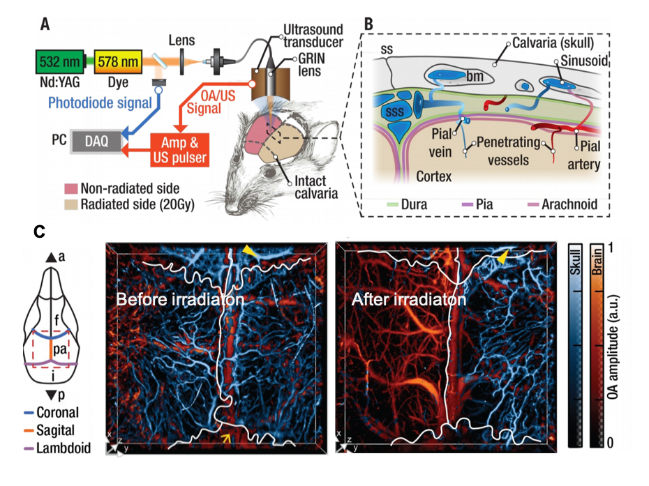
Various imaging methods have been established to overcome these limitations. Among them is optoacoustic ultrasound imaging, which has been pioneered and implemented for various biological questions by a handful of groups worldwide, including Prof. Daniel Razansky’s (Figure 1A, B). In the work presented here, Estrada et al (2) use optoacoustic ultrasound bio-microscopy to study radiation-induced damage in the skull bone marrow and microvasculature, following radiotherapy (Figure 1C).
The calvarian bone marrow is an important site for blood and immune cell generation and is maintained by the complex microvasculature network, composed by the calvarian sinusoids. Damage to this vascular network is clinically relevant for homeostasis, as the sinusoids provide an interface between the hematopoietically active bone marrow and the peripheral circulation.
The study sheds light on vascular changes upon radiotherapy, previously poorly understood, but which has important translational relevance, as radiotherapy is commonly used for cancer treatment.
Key findings
- In this work, Estrada et al used a hybrid imaging approach based on optoacoustic and ultrasound bio-microscopy. This approach allowed imaging over 6mm across the skull, with a spatial resolution of 12um and 30um in lateral and axial dimensions.
- The work introduces a novel segmentation method that allows differentiation of the calvarian vasculature based on its elastic and structural properties. This enabled distinguishing this microvascular network from the cerebral one.
- At 11 weeks following radiotherapy on half of the skull, key differences were noted between radiated and irradiated hemispheres: the sinusoidal vascular network in the calvarium remained intact only in the non-radiated hemisphere, while vasculature in the irradiated hemisphere (20 Gy) did not develop (Figure 1C).
- Quantitative analysis of vascular changes showed not only a decrease in number of vessels in irradiated hemispheres, but also a decrease in the number of branch points detected in the vascular network, suggesting altogether a loss of complexity in the vascular network.
Open questions and what I like about this paper
I liked this paper because of the usefulness of the tools developed by the authors, and the translationally relevant aspect of the findings, to vascular, and cancer research.
- Given the non-invasive nature of the method, and since you mention previous links to radiation-induced long-term cognitive disability, would it be possible in the future, to combine this method with other imaging platforms such as functional MRI, to study the direct link of angiogenesis inhibition, with collateral effects of irradiation?
- In the past, your lab has generated hybrid systems combining fluorescent and optoacoustic methods. Could you combine your findings at mesoscopic level with fluorescence methods to image hematopoietic niche changes, and immune cell dynamics derived from damage to the calvarian microvasclature after irradiation?
- Your study focused on the effects of irradiation on vascular remodelling, and you discuss effects in the context of cancer treatment. Your hybrid imaging method to visualize large areas of the skull, and to differentiate between cerebral and calvarian bone marrow could be applied to study other pathologies where changes to both compartments are induced. Have you considered applying this to other research topics?
- Given the possibility to differentiate calvarian from cerebral vasculature, is it also possible to differentiate between other types of vasculature, to map them and to study the interactions between them? Is this tool limited to the skull, or can it be applied to other organs as well?
References
- Ntziachristos V, Going deeper than microscopy: the optical imaging frontier in biology, Nat Methods, 2010, 7(8):603-614. doi: 10.1038/nmeth.1483.
- Estrada H, Rebling J, Sievert W, Hladik D, Hofmann U, Gottschalk S,Tapio S, Multhoff G, Razansky D, Intravital optoacoustic ultrasound bio-microscopy reveals radiation-inhibited skull angiogenesis, bioRxiv, 2019, doi: https://doi.org/10.1101/500017
doi: https://doi.org/10.1242/prelights.10644
Read preprint (2 votes)
 Have your say
Sign up to customise the site to your preferences and to receive alerts
Register hereAlso in the biophysics category:
Mechanically-induced Septin Networks Protect Nuclear Integrity
Filipe Nunes Vicente
Loss of Sun2 ablates nuclear mechanosensing-driven extracellular matrix production and mitigates lung fibrosis
Beth Chopak
Shape independent fluidisation in epithelial monolayers
Sindhu Muthukrishnan
Also in the cancer biology category:
Nature-Inspired Nanoparticle Adiposomes Enable Targeted Delivery of Hydrophobic Drug for Anti-Cancer Treatment
Elizabeth Pyman
Targeting CXADR-mediated AKT signaling suppresses tumorigenesis and enhances chemotherapy efficacy in Ewing sarcoma
TheLangeLab et al.
Dual inhibition of GTP-bound (ON) and GDP-bound (OFF) KRASG12C suppresses PI3Kα and leads to potent tumor inhibition
Luis Luna Ramírez
Also in the pathology category:
Behavioral characteristics of an extremely old rhesus macaque in a zoo: Dementia-like symptoms and implications for quality of life of geriatric animals
Stefan Friedrich Wirth
EBV reprograms autoreactive anti-CNS B cells as antigen presenting cells in multiple sclerosis
Léa Bastien et al.
Clinically reported covert cerebrovascular disease and risk of neurological disease: a whole-population cohort of 367,988 people using natural language processing
Rafidah Mumtahinah Chowdhury et al.
Also in the physiology category:
Inhibition of the gut ceramidase Asah2 decelerates the vertebrate ageing rate
Jeny Jose
Feeding and reproduction of a tropical coastal copepod across warming and copper gradients
Tina Nguyen
Resilience to cardiac aging in Greenland shark Somniosus microcephalus
Theodora Stougiannou
preLists in the biophysics category:
October in preprints – DevBio & Stem cell biology
Each month, preLighters with expertise across developmental and stem cell biology nominate a few recent developmental and stem cell biology (and related) preprints they’re excited about and explain in a single paragraph why. Short, snappy picks from working scientists — a quick way to spot fresh ideas, bold methods and papers worth reading in full. These preprints can all be found in the October preprint list published on the Node.
| List by | Deevitha Balasubramanian et al. |
October in preprints – Cell biology edition
Different preLighters, with expertise across cell biology, have worked together to create this preprint reading list for researchers with an interest in cell biology. This month, most picks fall under (1) Cell organelles and organisation, followed by (2) Mechanosignaling and mechanotransduction, (3) Cell cycle and division and (4) Cell migration
| List by | Matthew Davies et al. |
March in preprints – the CellBio edition
A group of preLighters, with expertise in different areas of cell biology, have worked together to create this preprint reading lists for researchers with an interest in cell biology. This month, categories include: 1) cancer biology 2) cell migration 3) cell organelles and organisation 4) cell signalling and mechanosensing 5) genetics and genomics 6) other
| List by | Girish Kale et al. |
Biologists @ 100 conference preList
This preList aims to capture all preprints being discussed at the Biologists @100 conference in Liverpool, UK, either as part of the poster sessions or the (flash/short/full-length) talks.
| List by | Reinier Prosee, Jonathan Townson |
February in preprints – the CellBio edition
A group of preLighters, with expertise in different areas of cell biology, have worked together to create this preprint reading lists for researchers with an interest in cell biology. This month, categories include: 1) biochemistry and cell metabolism 2) cell organelles and organisation 3) cell signalling, migration and mechanosensing
| List by | Barbora Knotkova et al. |
preLights peer support – preprints of interest
This is a preprint repository to organise the preprints and preLights covered through the 'preLights peer support' initiative.
| List by | preLights peer support |
66th Biophysical Society Annual Meeting, 2022
Preprints presented at the 66th BPS Annual Meeting, Feb 19 - 23, 2022 (The below list is not exhaustive and the preprints are listed in no particular order.)
| List by | Soni Mohapatra |
EMBL Synthetic Morphogenesis: From Gene Circuits to Tissue Architecture (2021)
A list of preprints mentioned at the #EESmorphoG virtual meeting in 2021.
| List by | Alex Eve |
Biophysical Society Meeting 2020
Some preprints presented at the Biophysical Society Meeting 2020 in San Diego, USA.
| List by | Tessa Sinnige |
ASCB EMBO Annual Meeting 2019
A collection of preprints presented at the 2019 ASCB EMBO Meeting in Washington, DC (December 7-11)
| List by | Madhuja Samaddar et al. |
EMBL Seeing is Believing – Imaging the Molecular Processes of Life
Preprints discussed at the 2019 edition of Seeing is Believing, at EMBL Heidelberg from the 9th-12th October 2019
| List by | Dey Lab |
Biomolecular NMR
Preprints related to the application and development of biomolecular NMR spectroscopy
| List by | Reid Alderson |
Biophysical Society Annual Meeting 2019
Few of the preprints that were discussed in the recent BPS annual meeting at Baltimore, USA
| List by | Joseph Jose Thottacherry |
Also in the cancer biology category:
BSDB Spring Meeting: Molecules to Morphogenesis
The British Society for Developmental Biology (BSDB) Spring Meeting Molecules to Morphogenesis was held from 23–26 March 2026 at the University of Warwick (UK). This meeting brought together a vibrant community of researchers to discuss how molecular mechanisms are integrated across scales to drive morphogenesis, spanning diverse model systems and approaches. This preList contains preprints by presenters from the talk and poster sessions at the meeting. Please do get in touch at preLights@biologists.com if you notice any relevant preprints that we may have missed.
| List by | Ingrid Tsang |
October in preprints – Cell biology edition
Different preLighters, with expertise across cell biology, have worked together to create this preprint reading list for researchers with an interest in cell biology. This month, most picks fall under (1) Cell organelles and organisation, followed by (2) Mechanosignaling and mechanotransduction, (3) Cell cycle and division and (4) Cell migration
| List by | Matthew Davies et al. |
September in preprints – Cell biology edition
A group of preLighters, with expertise in different areas of cell biology, have worked together to create this preprint reading list. This month, categories include: (1) Cell organelles and organisation, (2) Cell signalling and mechanosensing, (3) Cell metabolism, (4) Cell cycle and division, (5) Cell migration
| List by | Sristilekha Nath et al. |
July in preprints – the CellBio edition
A group of preLighters, with expertise in different areas of cell biology, have worked together to create this preprint reading lists for researchers with an interest in cell biology. This month, categories include: (1) Cell Signalling and Mechanosensing (2) Cell Cycle and Division (3) Cell Migration and Cytoskeleton (4) Cancer Biology (5) Cell Organelles and Organisation
| List by | Girish Kale et al. |
June in preprints – the CellBio edition
A group of preLighters, with expertise in different areas of cell biology, have worked together to create this preprint reading lists for researchers with an interest in cell biology. This month, categories include: (1) Cell organelles and organisation (2) Cell signaling and mechanosensation (3) Genetics/gene expression (4) Biochemistry (5) Cytoskeleton
| List by | Barbora Knotkova et al. |
May in preprints – the CellBio edition
A group of preLighters, with expertise in different areas of cell biology, have worked together to create this preprint reading lists for researchers with an interest in cell biology. This month, categories include: 1) Biochemistry/metabolism 2) Cancer cell Biology 3) Cell adhesion, migration and cytoskeleton 4) Cell organelles and organisation 5) Cell signalling and 6) Genetics
| List by | Barbora Knotkova et al. |
Keystone Symposium – Metabolic and Nutritional Control of Development and Cell Fate
This preList contains preprints discussed during the Metabolic and Nutritional Control of Development and Cell Fate Keystone Symposia. This conference was organized by Lydia Finley and Ralph J. DeBerardinis and held in the Wylie Center and Tupper Manor at Endicott College, Beverly, MA, United States from May 7th to 9th 2025. This meeting marked the first in-person gathering of leading researchers exploring how metabolism influences development, including processes like cell fate, tissue patterning, and organ function, through nutrient availability and metabolic regulation. By integrating modern metabolic tools with genetic and epidemiological insights across model organisms, this event highlighted key mechanisms and identified open questions to advance the emerging field of developmental metabolism.
| List by | Virginia Savy, Martin Estermann |
April in preprints – the CellBio edition
A group of preLighters, with expertise in different areas of cell biology, have worked together to create this preprint reading lists for researchers with an interest in cell biology. This month, categories include: 1) biochemistry/metabolism 2) cell cycle and division 3) cell organelles and organisation 4) cell signalling and mechanosensing 5) (epi)genetics
| List by | Vibha SINGH et al. |
March in preprints – the CellBio edition
A group of preLighters, with expertise in different areas of cell biology, have worked together to create this preprint reading lists for researchers with an interest in cell biology. This month, categories include: 1) cancer biology 2) cell migration 3) cell organelles and organisation 4) cell signalling and mechanosensing 5) genetics and genomics 6) other
| List by | Girish Kale et al. |
Biologists @ 100 conference preList
This preList aims to capture all preprints being discussed at the Biologists @100 conference in Liverpool, UK, either as part of the poster sessions or the (flash/short/full-length) talks.
| List by | Reinier Prosee, Jonathan Townson |
February in preprints – the CellBio edition
A group of preLighters, with expertise in different areas of cell biology, have worked together to create this preprint reading lists for researchers with an interest in cell biology. This month, categories include: 1) biochemistry and cell metabolism 2) cell organelles and organisation 3) cell signalling, migration and mechanosensing
| List by | Barbora Knotkova et al. |
BSCB-Biochemical Society 2024 Cell Migration meeting
This preList features preprints that were discussed and presented during the BSCB-Biochemical Society 2024 Cell Migration meeting in Birmingham, UK in April 2024. Kindly put together by Sara Morais da Silva, Reviews Editor at Journal of Cell Science.
| List by | Reinier Prosee |
CSHL 87th Symposium: Stem Cells
Preprints mentioned by speakers at the #CSHLsymp23
| List by | Alex Eve |
Journal of Cell Science meeting ‘Imaging Cell Dynamics’
This preList highlights the preprints discussed at the JCS meeting 'Imaging Cell Dynamics'. The meeting was held from 14 - 17 May 2023 in Lisbon, Portugal and was organised by Erika Holzbaur, Jennifer Lippincott-Schwartz, Rob Parton and Michael Way.
| List by | Helen Zenner |
CellBio 2022 – An ASCB/EMBO Meeting
This preLists features preprints that were discussed and presented during the CellBio 2022 meeting in Washington, DC in December 2022.
| List by | Nadja Hümpfer et al. |
Fibroblasts
The advances in fibroblast biology preList explores the recent discoveries and preprints of the fibroblast world. Get ready to immerse yourself with this list created for fibroblasts aficionados and lovers, and beyond. Here, my goal is to include preprints of fibroblast biology, heterogeneity, fate, extracellular matrix, behavior, topography, single-cell atlases, spatial transcriptomics, and their matrix!
| List by | Osvaldo Contreras |
Single Cell Biology 2020
A list of preprints mentioned at the Wellcome Genome Campus Single Cell Biology 2020 meeting.
| List by | Alex Eve |
ASCB EMBO Annual Meeting 2019
A collection of preprints presented at the 2019 ASCB EMBO Meeting in Washington, DC (December 7-11)
| List by | Madhuja Samaddar et al. |
Lung Disease and Regeneration
This preprint list compiles highlights from the field of lung biology.
| List by | Rob Hynds |
Anticancer agents: Discovery and clinical use
Preprints that describe the discovery of anticancer agents and their clinical use. Includes both small molecules and macromolecules like biologics.
| List by | Zhang-He Goh |
Also in the pathology category:
preLighters’ choice – Handpicked DevBio preprints
preLighters with expertise across developmental and stem cell biology have nominated a few developmental biology (and related) preprints they’re excited about and explain in a few paragraph why. Concise preprint highlights, prepared by the preLighter community – a quick way to spot upcoming trends, new methods and fresh ideas.
| List by | Theodora Stougiannou et al. |
October in preprints – DevBio & Stem cell biology
Each month, preLighters with expertise across developmental and stem cell biology nominate a few recent developmental and stem cell biology (and related) preprints they’re excited about and explain in a single paragraph why. Short, snappy picks from working scientists — a quick way to spot fresh ideas, bold methods and papers worth reading in full. These preprints can all be found in the October preprint list published on the Node.
| List by | Deevitha Balasubramanian et al. |
October in preprints – Cell biology edition
Different preLighters, with expertise across cell biology, have worked together to create this preprint reading list for researchers with an interest in cell biology. This month, most picks fall under (1) Cell organelles and organisation, followed by (2) Mechanosignaling and mechanotransduction, (3) Cell cycle and division and (4) Cell migration
| List by | Matthew Davies et al. |
Fibroblasts
The advances in fibroblast biology preList explores the recent discoveries and preprints of the fibroblast world. Get ready to immerse yourself with this list created for fibroblasts aficionados and lovers, and beyond. Here, my goal is to include preprints of fibroblast biology, heterogeneity, fate, extracellular matrix, behavior, topography, single-cell atlases, spatial transcriptomics, and their matrix!
| List by | Osvaldo Contreras |
ECFG15 – Fungal biology
Preprints presented at 15th European Conference on Fungal Genetics 17-20 February 2020 Rome
| List by | Hiral Shah |
COVID-19 / SARS-CoV-2 preprints
List of important preprints dealing with the ongoing coronavirus outbreak. See http://covidpreprints.com for additional resources and timeline, and https://connect.biorxiv.org/relate/content/181 for full list of bioRxiv and medRxiv preprints on this topic
| List by | Dey Lab, Zhang-He Goh |
1
Cellular metabolism
A curated list of preprints related to cellular metabolism at Biorxiv by Pablo Ranea Robles from the Prelights community. Special interest on lipid metabolism, peroxisomes and mitochondria.
| List by | Pablo Ranea Robles |
Also in the physiology category:
Developmental regulation: molecular and ecological niches
This conference was held at the Station Biologique de Roscoff (France) and brought together researchers exploring how diverse niche environments shape developmental processes across scales. Spanning topics from ecological and metabolic influences to signalling networks, mechanics and gene regulation, the meeting highlighted the interplay between intrinsic and extrinsic factors in controlling cell fate and tissue organisation. This preList gathers preprints discussed by speakers and poster presenters during the meeting. Please do get in touch at preLights@biologists.com if you notice any relevant preprints that we may have missed.
| List by | Ingrid Tsang |
preLighters’ choice – Handpicked DevBio preprints
preLighters with expertise across developmental and stem cell biology have nominated a few developmental biology (and related) preprints they’re excited about and explain in a few paragraph why. Concise preprint highlights, prepared by the preLighter community – a quick way to spot upcoming trends, new methods and fresh ideas.
| List by | Theodora Stougiannou et al. |
Keystone Symposium on Stem Cell Models in Embryology 2026
The Keystone Symposium on Stem Cell Models in Embryology, 2026, was organised by Jun Wu (UT Southwestern), Jianping Fu (University of Michigan) and Miki Ebisuya (TU Dresden) and held at Asilomar Conference Grounds in California (US). The meeting discussed recent advances made in establishing stem-cell-based embryo models, what fundamental insights into developmental processes have been gleaned from them, as well as how they are beginning to be applied more widely. This prelist contains preprints by presenters at the talk and poster sessions at the conference, which our Reviews Editor in attendance spotted. Please do reach out to preLights@biologists.com if you notice any that we’ve missed.
| List by | Ingrid Tsang |
October in preprints – DevBio & Stem cell biology
Each month, preLighters with expertise across developmental and stem cell biology nominate a few recent developmental and stem cell biology (and related) preprints they’re excited about and explain in a single paragraph why. Short, snappy picks from working scientists — a quick way to spot fresh ideas, bold methods and papers worth reading in full. These preprints can all be found in the October preprint list published on the Node.
| List by | Deevitha Balasubramanian et al. |
Biologists @ 100 conference preList
This preList aims to capture all preprints being discussed at the Biologists @100 conference in Liverpool, UK, either as part of the poster sessions or the (flash/short/full-length) talks.
| List by | Reinier Prosee, Jonathan Townson |
Fibroblasts
The advances in fibroblast biology preList explores the recent discoveries and preprints of the fibroblast world. Get ready to immerse yourself with this list created for fibroblasts aficionados and lovers, and beyond. Here, my goal is to include preprints of fibroblast biology, heterogeneity, fate, extracellular matrix, behavior, topography, single-cell atlases, spatial transcriptomics, and their matrix!
| List by | Osvaldo Contreras |
FENS 2020
A collection of preprints presented during the virtual meeting of the Federation of European Neuroscience Societies (FENS) in 2020
| List by | Ana Dorrego-Rivas |
TAGC 2020
Preprints recently presented at the virtual Allied Genetics Conference, April 22-26, 2020. #TAGC20
| List by | Maiko Kitaoka et al. |
Autophagy
Preprints on autophagy and lysosomal degradation and its role in neurodegeneration and disease. Includes molecular mechanisms, upstream signalling and regulation as well as studies on pharmaceutical interventions to upregulate the process.
| List by | Sandra Malmgren Hill |
6 years
Hector Estrada
Dear Mariana,
Thank you for your interest in our work.
Answering your open questions:
1.- Given the non-invasive nature of the method, and since you mention previous links to radiation-induced long-term cognitive disability, would it be possible in the future, to combine this method with other imaging platforms such as functional MRI, to study the direct link of angiogenesis inhibition, with collateral effects of irradiation?
At the beginning of our study we knew that imaging bone vasculature in-vivo was challenging. We learned that it required at least two complementary techniques (optoacoustics and ultrasound) to tackle the problem. Then, in order to answer more involved biological questions such as how angiongenesis inhibition is linked to other collateral effects of irradiation, we certainly need to include other imaging platforms that could offer complementary information. We think it is technically feasible.
2.- In the past, your lab has generated hybrid systems combining fluorescent and optoacoustic methods. Could you combine your findings at mesoscopic level with fluorescence methods to image hematopoietic niche changes, and immune cell dynamics derived from damage to the calvarian microvasclature after irradiation?
In the current article we show what can be done with intrinsic contrast. Combining it with fluorescence imaging and sub-cellular resolution would result in a very exciting platform. Such a multiscale system can rely on optoacoustic and ultrasound to observe large scale effects of radiation and then zoom-in at an interesting spot to look at the details of cell dynamics using fluorescence labels. We also hope that people developing optoacoustic labels could soon help us reaching even further.
3.- Your study focused on the effects of irradiation on vascular remodelling, and you discuss effects in the context of cancer treatment. Your hybrid imaging method to visualize large areas of the skull, and to differentiate between cerebral and calvarian bone marrow could be applied to study other pathologies where changes to both compartments are induced. Have you considered applying this to other research topics?
We are certainly looking forward to using our method to study other pathologies that could affect the skull/brain interface. One could think of tumors and some other bone lesions.
4.- Given the possibility to differentiate calvarian from cerebral vasculature, is it also possible to differentiate between other types of vasculature, to map them and to study the interactions between them? Is this tool limited to the skull, or can it be applied to other organs as well?
It would be great if we could observe the lymphatic system as well. In order to do that we need either an extrinsic contrast or combine our method with fluorescence imaging.
Imaging bone vasculature in-vivo is technically very challenging, not only for optical methods. Ultrasound waves are greatly distorted when propagating through bone; in fact, most of the wave is reflected back. High resolution optoacoustic systems developed to image the mouse brain can only image very young mice or need to perform a craniotomy. Our method extends the applicability of high resolution optoacoustics and shows how to handle the skull and its vasculature properly. In addition, the skull is an excellent window to observe what happens inside the bone and it is right next to the brain, which is the most interesting organ. There are some examples in the literature that show segmentation of optoacoustic images in bone-less regions of the body. In our opinion, the skull is pivotal because it allows us to observe interesting biological processes and also paves the way for exploring other regions of the body where bones pose a problem for imaging.