






Choreographed morphogenetic events underlie early foregut development in the mouse embryo
Posted on: 23 September 2026
Preprint posted on 24 August 2026

Categories: developmental biology
Why I chose this preprint
The anterior embryo is one of the best-mapped regions in developmental biology at the molecular level — but the large-scale morphogenetic movements that actually build its geometry, and especially how multiple tissues coordinate to produce them, remain largely overlooked. This preprint addresses this gap directly: the long-term 3D live-imaging dataset presented here is an amazing resource for studying how these dramatic, coordinated shape changes are orchestrated. The authors took a good first step in identifying and quantitatively describing the stereotypical sequence of foregut involution and began testing several candidate processes that might contribute to it. Still, many of the driving mechanisms remain speculative. I hope highlighting this work brings it wider visibility and draws more attention to these relatively underexplored areas in embryonic morphogenesis.
Background
In the mouse embryo, organogenesis of the brain, heart, and gut is initiated at the anterior end during embryonic days 7.5 to 8.5 (E7.5–E8.5): the cranial neural plate elevates and generates the forebrain; the cardiac crescent coalesces to form the linear heart tube; and the anterior endoderm folds inward to form the foregut pocket.
This preprint focuses on foregut involution. At E7.5, the anterior surface endoderm is a salt-and-pepper mosaic of 1) embryonic visceral endoderm (emVE) cells derived from the primitive endoderm and 2) definitive endoderm (DE) cells derived from the epiblast, which ingress through the primitive streak (PS) during gastrulation and intercalate into the emVE. This squamous endodermal sheet rapidly involutes and extends inward to give rise to the anterior gastrointestinal organs.
Key Findings
1. Long-term live imaging of E7.5 mouse embryos reveals a stereotyped sequence of foregut involution
Using lightsheet and spinning-disc microscopy, the authors live-imaged the full process of foregut involution from E7.5 to ~E8 and described its morphological sequence.
Quantitatively, spherical-harmonics-based analysis confirmed that the involution process is highly consistent between embryos.
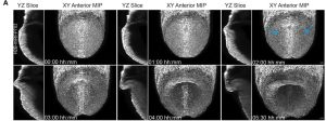
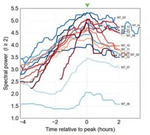
Fig. 1 Long-term live imaging of E7.5 mouse embryos using light-sheet microscopy captured a stereotypical sequence of involution. Figure adapted from the preprint where it is available under a CC-BY-NC 4.0 International license.
2. Actomyosin contractility appears not required for initiating involution
The authors reported intense cortical actin condensation along the central midline of the embryo, especially in the forming pocket.
However, immunostaining did not show high levels of phosphorylated myosin light chain accompanying this actin enrichment. In addition, treatment with the myosin inhibitor blebbistatin or the ROCK inhibitor Rockout did not prevent the initiation of folding.
3. Foregut involution is spatiotemporally correlated with a peak of cell death in the embryonic visceral endoderm (emVE) lineage, yet is not dependent on it
Tracking in the live-imaging data showed a peak of cell death in the anterior endoderm coinciding with foregut involution. More detailed inspection showed that dying cells assemble a contractile actomyosin ring at their apex, and apoptotic bodies are subsequently extruded bidirectionally toward both the apical and basal sides, with a basal bias.
TUNEL assays and cleaved-caspase-3 (cCasp3) immunostaining in emVE-labelled embryos confirmed that these cell deaths are largely restricted to emVE lineage cells.
Treatment with a caspase inhibitor or emVE-specific overexpression of the anti-apoptotic factor BCL-2 effectively blocked emVE cell death and resulted in the formation of multilayered emVE cell aggregates across the anterior endoderm; however, foregut involution was not greatly disrupted.
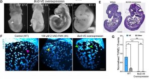
Fig. 2 Blocking emVE-specific cell deaths resulted in formation of emVE cell aggregates but did not significantly disrupt foregut morphogenesis. Figure adapted from the preprint where it is available under a CC-BY-NC 4.0 International license.
4. Transcriptional profiles show clear distinctions between emVE/DE lineages and anterior/posterior positions
The authors performed bulk RNA-seq on embryos carrying both emVE (Afp-kGFP) and pan-endoderm (Foxa2-eGFP) markers, which allowed identification and FACS sorting of emVE and DE cells.
PCA separated emVE and DE cells into distinct clusters and further resolved anterior and posterior populations within both lineages.
Among the differentially expressed genes, the authors highlighted upregulation of morphogenesis- and cell-adhesion-related genes in anterior DE, and downregulation of cell-cycle genes in anterior emVE.
5. Detailed characterization of invagination defects following VE-specific Bmp2 deletion
Bmp2 knockout in VE cells has been reported to result in anterior disorganization. Bulk RNA-seq confirmed Bmp2 expression in emVE, as well as the presence of BMP receptors and canonical SMADs, with Acvr1/Bmpr2 and Smad5 showing positional and stage-dependent correlations, respectively, potentially linking them to the anterior-specific function of BMP2.
The authors live-imaged Bmp2 VE-KO embryos, showing that the majority managed to form a shallow foregut pocket or indentation, but at an aberrant position and with delayed timing. Subsequent ventral folding lost central symmetry and was accompanied by aberrant folding and buckling of the neural headfolds.
The authors noted that the position of the neural headfolds still correlated with the position of lateral endodermal involutions, and that failed central foregut pocket formation was also concurrent with perturbed positioning of the heart field. They therefore proposed that these structures are coupled and act as a single mechanical unit.
In addition, notochord morphogenesis was severely perturbed in Bmp2 VE-KO embryos, with the notochord becoming wider and showing compromised continuity. Spatial restriction of emVE apoptosis was also lost in Bmp2 VE-KO embryos, with cell death occurring broadly across the anterior endoderm rather than being restricted to the central proximal region.
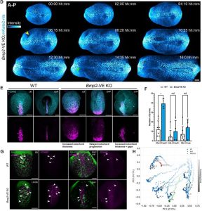
Fig. 3 Bmp2 VE-KO embryos showed aberrant positioning and progression of involution, accompanied by disruption of notochord morphology and widespread cell deaths across endoderm. Figure adapted from the preprint where it is available under a CC-BY-NC 4.0 International license.
Questions to the authors
- I find it rather surprising that compromising actomyosin contractility does not prevent the initiation of folding. Do you have any hypotheses about what the key driver of foregut invagination might be? For example, do the deformation patterns visible in your live-imaging data provide any clues about the tissue-level forces or strains involved?
- The tight correlation between the neural headfolds and foregut is very interesting. Do you have any indications of how the endoderm and ectoderm might be mechanically coupled during foregut involution — for example, through ECM distribution/architecture or coordinated versus relative tissue movements? Based on the morphogenetic dynamics you observe, which tissue do you think is most likely to provide the primary driving force?
- Avian and reptile embryos also undergo large-scale anterior folding that shapes the forebrain, heart, and foregut at roughly equivalent developmental stages. However, these embryos begin from a flat, multilayer disc shape, in contrast to the cup-shaped mouse embryo. Do you think the underlying mechanisms coordinating these morphogenetic movements are likely to be conserved across species, or might the different initial geometries require fundamentally different mechanical strategies?
doi: https://doi.org/10.1242/prelights.44928
Read preprintSign up to customise the site to your preferences and to receive alerts
Register hereAlso in the developmental biology category:
'Preprints by preLighters': A novel reaction-diffusion architecture for engineering self-organized patterns in mammalian cells
Reinier Prosee, Benjamin Swedlund

Clonal embeddings allow exploratory analysis of lineage-resolved single-cell data
Marine Secchi

E-cadherin clustering as a regulator of morphogenesis
Girish Kale, preLights peer support

preLists in the developmental biology category:
‘In preprints’ from Development 2026
A list of the preprints featured in Development's 'In preprints' articles, as part of the new Preprint Editors initiative
| List by | Clarisse Brunet Avalos et al. |
Sainsbury Laboratory Symposium 2026
The Sainsbury Laboratory Symposium 2026, Shaping Life: Mechanisms of Morphogenesis, brought together researchers exploring the cellular, genetic and mechanical processes that shape plant form. This preList highlights preprints that were discussed or mentioned at the meeting. If you notice any missing preprints, please contact us at preLights@biologists.com
| List by | Ingrid Tsang |
Santa Cruz Developmental Biology Meeting 2026
This preList brings together a selection of preprints discussed at the Santa Cruz Developmental Biology Meeting 2026, held at UC Santa Cruz with the theme “Biomedicine, Biomechanics, and the Biosphere.” Reflecting the meeting’s broad scientific scope, from development and regeneration to evolution, tissue mechanics, and emerging technologies, this curated collection showcases work that contributed to conversations throughout the meeting.
| List by | Ingrid Tsang |
Developmental regulation: molecular and ecological niches
This conference was held at the Station Biologique de Roscoff (France) and brought together researchers exploring how diverse niche environments shape developmental processes across scales. Spanning topics from ecological and metabolic influences to signalling networks, mechanics and gene regulation, the meeting highlighted the interplay between intrinsic and extrinsic factors in controlling cell fate and tissue organisation. This preList gathers preprints discussed by speakers and poster presenters during the meeting. Please do get in touch at preLights@biologists.com if you notice any relevant preprints that we may have missed.
| List by | Ingrid Tsang |
preLighters’ choice – Handpicked DevBio preprints
preLighters with expertise across developmental and stem cell biology have nominated a few developmental biology (and related) preprints they’re excited about and explain in a few paragraph why. Concise preprint highlights, prepared by the preLighter community – a quick way to spot upcoming trends, new methods and fresh ideas.
| List by | Theodora Stougiannou et al. |
BSDB Spring Meeting: Molecules to Morphogenesis
The British Society for Developmental Biology (BSDB) Spring Meeting Molecules to Morphogenesis was held from 23–26 March 2026 at the University of Warwick (UK). This meeting brought together a vibrant community of researchers to discuss how molecular mechanisms are integrated across scales to drive morphogenesis, spanning diverse model systems and approaches. This preList contains preprints by presenters from the talk and poster sessions at the meeting. Please do get in touch at preLights@biologists.com if you notice any relevant preprints that we may have missed.
| List by | Ingrid Tsang |
Keystone Symposium on Stem Cell Models in Embryology 2026
The Keystone Symposium on Stem Cell Models in Embryology, 2026, was organised by Jun Wu (UT Southwestern), Jianping Fu (University of Michigan) and Miki Ebisuya (TU Dresden) and held at Asilomar Conference Grounds in California (US). The meeting discussed recent advances made in establishing stem-cell-based embryo models, what fundamental insights into developmental processes have been gleaned from them, as well as how they are beginning to be applied more widely. This prelist contains preprints by presenters at the talk and poster sessions at the conference, which our Reviews Editor in attendance spotted. Please do reach out to preLights@biologists.com if you notice any that we’ve missed.
| List by | Ingrid Tsang |
November in preprints – DevBio & Stem cell biology
preLighters with expertise across developmental and stem cell biology have nominated a few developmental and stem cell biology (and related) preprints posted in November they’re excited about and explain in a single paragraph why. Concise preprint highlights, prepared by the preLighter community – a quick way to spot upcoming trends, new methods and fresh ideas.
| List by | Aline Grata et al. |
October in preprints – DevBio & Stem cell biology
Each month, preLighters with expertise across developmental and stem cell biology nominate a few recent developmental and stem cell biology (and related) preprints they’re excited about and explain in a single paragraph why. Short, snappy picks from working scientists — a quick way to spot fresh ideas, bold methods and papers worth reading in full. These preprints can all be found in the October preprint list published on the Node.
| List by | Deevitha Balasubramanian et al. |
October in preprints – Cell biology edition
Different preLighters, with expertise across cell biology, have worked together to create this preprint reading list for researchers with an interest in cell biology. This month, most picks fall under (1) Cell organelles and organisation, followed by (2) Mechanosignaling and mechanotransduction, (3) Cell cycle and division and (4) Cell migration
| List by | Matthew Davies et al. |
June in preprints – the CellBio edition
A group of preLighters, with expertise in different areas of cell biology, have worked together to create this preprint reading lists for researchers with an interest in cell biology. This month, categories include: (1) Cell organelles and organisation (2) Cell signaling and mechanosensation (3) Genetics/gene expression (4) Biochemistry (5) Cytoskeleton
| List by | Barbora Knotkova et al. |
Keystone Symposium – Metabolic and Nutritional Control of Development and Cell Fate
This preList contains preprints discussed during the Metabolic and Nutritional Control of Development and Cell Fate Keystone Symposia. This conference was organized by Lydia Finley and Ralph J. DeBerardinis and held in the Wylie Center and Tupper Manor at Endicott College, Beverly, MA, United States from May 7th to 9th 2025. This meeting marked the first in-person gathering of leading researchers exploring how metabolism influences development, including processes like cell fate, tissue patterning, and organ function, through nutrient availability and metabolic regulation. By integrating modern metabolic tools with genetic and epidemiological insights across model organisms, this event highlighted key mechanisms and identified open questions to advance the emerging field of developmental metabolism.
| List by | Virginia Savy, Martin Estermann |
Biologists @ 100 conference preList
This preList aims to capture all preprints being discussed at the Biologists @100 conference in Liverpool, UK, either as part of the poster sessions or the (flash/short/full-length) talks.
| List by | Reinier Prosee, Jonathan Townson |
BSDB/GenSoc Spring Meeting 2024
A list of preprints highlighted at the British Society for Developmental Biology and Genetics Society joint Spring meeting 2024 at Warwick, UK.
| List by | Joyce Yu, Katherine Brown |
GfE/ DSDB meeting 2024
This preList highlights the preprints discussed at the 2024 joint German and Dutch developmental biology societies meeting that took place in March 2024 in Osnabrück, Germany.
| List by | Joyce Yu |
‘In preprints’ from Development 2022-2023
A list of the preprints featured in Development's 'In preprints' articles between 2022-2023
| List by | Alex Eve, Katherine Brown |
preLights peer support – preprints of interest
This is a preprint repository to organise the preprints and preLights covered through the 'preLights peer support' initiative.
| List by | preLights peer support |
The Society for Developmental Biology 82nd Annual Meeting
This preList is made up of the preprints discussed during the Society for Developmental Biology 82nd Annual Meeting that took place in Chicago in July 2023.
| List by | Joyce Yu, Katherine Brown |
CSHL 87th Symposium: Stem Cells
Preprints mentioned by speakers at the #CSHLsymp23
| List by | Alex Eve |
Journal of Cell Science meeting ‘Imaging Cell Dynamics’
This preList highlights the preprints discussed at the JCS meeting 'Imaging Cell Dynamics'. The meeting was held from 14 - 17 May 2023 in Lisbon, Portugal and was organised by Erika Holzbaur, Jennifer Lippincott-Schwartz, Rob Parton and Michael Way.
| List by | Helen Zenner |
9th International Symposium on the Biology of Vertebrate Sex Determination
This preList contains preprints discussed during the 9th International Symposium on the Biology of Vertebrate Sex Determination. This conference was held in Kona, Hawaii from April 17th to 21st 2023.
| List by | Martin Estermann |
Alumni picks – preLights 5th Birthday
This preList contains preprints that were picked and highlighted by preLights Alumni - an initiative that was set up to mark preLights 5th birthday. More entries will follow throughout February and March 2023.
| List by | Sergio Menchero et al. |
CellBio 2022 – An ASCB/EMBO Meeting
This preLists features preprints that were discussed and presented during the CellBio 2022 meeting in Washington, DC in December 2022.
| List by | Nadja Hümpfer et al. |
2nd Conference of the Visegrád Group Society for Developmental Biology
Preprints from the 2nd Conference of the Visegrád Group Society for Developmental Biology (2-5 September, 2021, Szeged, Hungary)
| List by | Nándor Lipták |
Fibroblasts
The advances in fibroblast biology preList explores the recent discoveries and preprints of the fibroblast world. Get ready to immerse yourself with this list created for fibroblasts aficionados and lovers, and beyond. Here, my goal is to include preprints of fibroblast biology, heterogeneity, fate, extracellular matrix, behavior, topography, single-cell atlases, spatial transcriptomics, and their matrix!
| List by | Osvaldo Contreras |
EMBL Synthetic Morphogenesis: From Gene Circuits to Tissue Architecture (2021)
A list of preprints mentioned at the #EESmorphoG virtual meeting in 2021.
| List by | Alex Eve |
EMBL Conference: From functional genomics to systems biology
Preprints presented at the virtual EMBL conference "from functional genomics and systems biology", 16-19 November 2020
| List by | Jesus Victorino |
Single Cell Biology 2020
A list of preprints mentioned at the Wellcome Genome Campus Single Cell Biology 2020 meeting.
| List by | Alex Eve |
Society for Developmental Biology 79th Annual Meeting
Preprints at SDB 2020
| List by | Irepan Salvador-Martinez, Martin Estermann |
FENS 2020
A collection of preprints presented during the virtual meeting of the Federation of European Neuroscience Societies (FENS) in 2020
| List by | Ana Dorrego-Rivas |
Planar Cell Polarity – PCP
This preList contains preprints about the latest findings on Planar Cell Polarity (PCP) in various model organisms at the molecular, cellular and tissue levels.
| List by | Ana Dorrego-Rivas |
Cell Polarity
Recent research from the field of cell polarity is summarized in this list of preprints. It comprises of studies focusing on various forms of cell polarity ranging from epithelial polarity, planar cell polarity to front-to-rear polarity.
| List by | Yamini Ravichandran |
TAGC 2020
Preprints recently presented at the virtual Allied Genetics Conference, April 22-26, 2020. #TAGC20
| List by | Maiko Kitaoka et al. |
3D Gastruloids
A curated list of preprints related to Gastruloids (in vitro models of early development obtained by 3D aggregation of embryonic cells). Updated until July 2021.
| List by | Paul Gerald L. Sanchez and Stefano Vianello |
ASCB EMBO Annual Meeting 2019
A collection of preprints presented at the 2019 ASCB EMBO Meeting in Washington, DC (December 7-11)
| List by | Madhuja Samaddar et al. |
EDBC Alicante 2019
Preprints presented at the European Developmental Biology Congress (EDBC) in Alicante, October 23-26 2019.
| List by | Sergio Menchero et al. |
EMBL Seeing is Believing – Imaging the Molecular Processes of Life
Preprints discussed at the 2019 edition of Seeing is Believing, at EMBL Heidelberg from the 9th-12th October 2019
| List by | Dey Lab |
SDB 78th Annual Meeting 2019
A curation of the preprints presented at the SDB meeting in Boston, July 26-30 2019. The preList will be updated throughout the duration of the meeting.
| List by | Alex Eve |
Lung Disease and Regeneration
This preprint list compiles highlights from the field of lung biology.
| List by | Rob Hynds |
Young Embryologist Network Conference 2019
Preprints presented at the Young Embryologist Network 2019 conference, 13 May, The Francis Crick Institute, London
| List by | Alex Eve |
Pattern formation during development
The aim of this preList is to integrate results about the mechanisms that govern patterning during development, from genes implicated in the processes to theoritical models of pattern formation in nature.
| List by | Alexa Sadier |
BSCB/BSDB Annual Meeting 2019
Preprints presented at the BSCB/BSDB Annual Meeting 2019
| List by | Dey Lab |
Zebrafish immunology
A compilation of cutting-edge research that uses the zebrafish as a model system to elucidate novel immunological mechanisms in health and disease.
| List by | Shikha Nayar |