


Plakoglobin is a mechanoresponsive regulator of naïve pluripotency
Posted on: 23 May 2022
Preprint posted on 16 March 2022
Plakoglobin to the rescue: how the vertebrate homologue of β-catenin senses mechanics during early embryogenesis to maintain naïve pluripotency.
Selected by Ilaria Di MeglioCategories: biophysics, developmental biology
Background
Physical and chemical stimuli from the external environment are key to development; they dictate the processes that transform a single cell into a complex multicellular organism (Heisenberg and Bellaiche, 2013). Even before implantation, during the earliest stages of embryogenesis, naive pluripotent cells must integrate these cues as they are directed towards specific cell fates (lineage specification) (Chan et al., 2017, Vining and Mooney, 2017). But we still lack full understanding of how mechanical signals contribute to this process. How are physical cues, like volumetric compression and spatial confinement, translated into transcriptional changes during naïve pluripotency? Which are the mechanosensitive genes involved?
In their study, Kohler and colleagues tackle these fundamental questions by studying the transcriptional and morphological development of naïve pluripotent embryonic stem cells (ESCs) encapsulated in 3D agarose microgels. Using this approach, they demonstrate that volumetric confinement affects pluripotency and show that expression of Plakoglobin, a vertebrate homologue of β-catenin, is required for naive pluripotency.
Key findings
Kohler et al encapsulated mouse ESCs inside spherical agarose microgels (Fig 1A). This system allows the authors to address their questions because mouse ESCs capture naïve pluripotency in vitro and microgels provide a 3D culture scaffold that mimics the volumetric confinement experienced by cells in the developing embryo. Once generated, microgels were cultured in either: self-renewal conditions in 2i/LIF medium, which sustains naïve pluripotency indefinitely; self-renewal conditions in serum/LIF, which supports a metastable state; or differentiating conditions in N2B27 medium to induce cell differentiation. The naïve (KLF4) and general (OCT4) pluripotency markers were used to confirm these conditions. Accordingly, pluripotency markers were expressed under self-renewal conditions, and upon transfer to differentiating conditions for 2 days, KLF4 and OCT4 were downregulated and absent, respectively. To assess the effects of 3D culture on ESCs, Kohler and colleagues utilized a reporter cell line, Rex1::GFPd2 (hereafter RGd2 cells) as a quantitative and real-time measure of naïve pluripotency in cells. Homogeneous GFP signal in 2i/LIF medium indicated naïve pluripotency whereas loss of expression in N2B27 indicated exit from naïve pluripotency. Thus, 3D agarose microgels are suitable for culture of naive pluripotent ESCs.
To assess how 3D microgels affect transcription, the authors compared gene expression data from naive ESCs in 3D microgels to naïve ESCs in 2D tissue culture. In addition to the global increase of naïve pluripotency markers in 3D, the most differentially expressed genes included components of the Wnt/β-catenin signaling pathways, notably Jup. The gene Jup encodes for the protein Plakoglobin, the vertebrate homologue of β-catenin. Analysis of the genes associated with cell-cell adherens junction (AJs), where β-catenin normally localizes, confirmed that Jup shows the strongest increase at AJs. Likewise, immunofluorescence analysis of the cell adhesion proteins at AJs confirmed that Plakoglobin localized to AJs in 3D microgels but not in 2D, whereas β-catenin was expressed in both but was higher in 2D (Fig 1B).
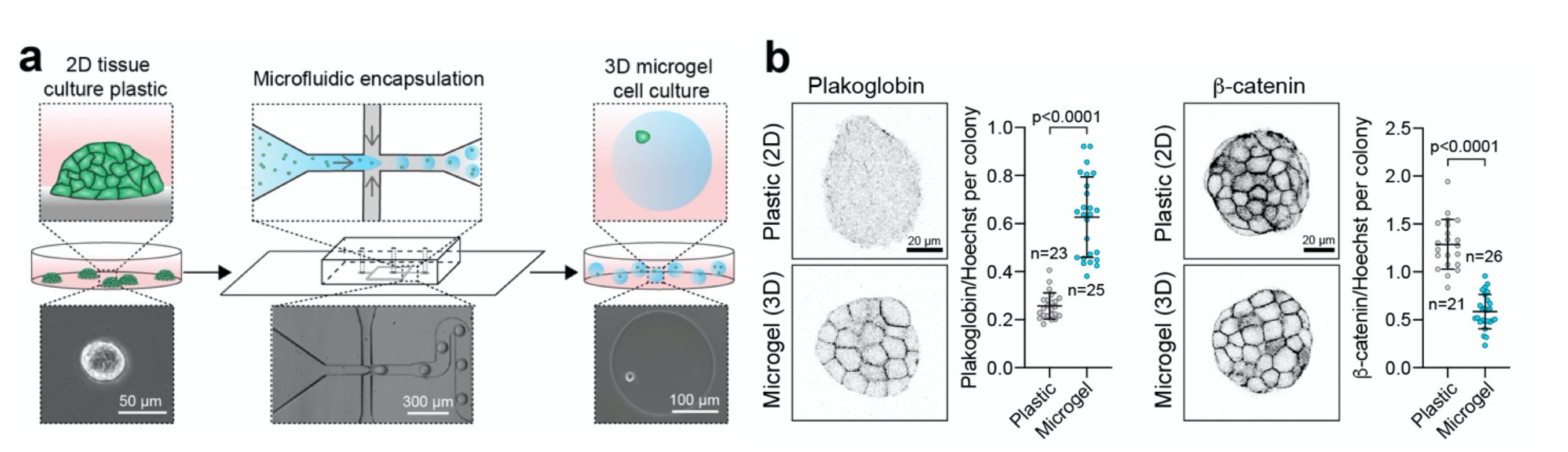
Figure 1 | ESC encapsulation in 3D microgels promotes expression of β-catenin homologue, Plakoglobin
a. Schematic of the microfluidic technique used to encapsulate ESCs in 3D microgels. b. Immunofluorescence images and quantification for Plakoglobin (left) and β-catenin (right) in 2D culture vs. 3D microgels.
Importantly, the authors also compared their findings to hanging drop cultures, where ESCs grow in a 3D environment without confinement. This culture system would allow them to confirm that the observed effects are due to volumetric confinement from 3D microgels and not just the 3D environment. They found that β-catenin was observed in both conditions, whereas Plakoglobin was upregulated only in 3D microgels. Even in the case where ESCs were released from 3D microgels after 48h and transferred to hanging drop cultures, Plakoglobin expression was lost. This confirmed that volumetric confinement generated by 3D microgels is required for Plakoglobin expression.
To corroborate the link between naïve pluripotency and Plakoglobin in vivo, Kohler and colleagues also analyzed human and mouse embryos. Consistent with their findings in naïve ESCs, they found high expression of Jup in the pre-implantation mouse embryo, when naïve pluripotency is established. At the protein level, the expression of Plakoglobin correlated with expansion of the blastocyst’s cavity. Although harder to assess in vivo, these observations suggest that an increase in pressure from expansion of the cavity may provide the mechanical signal that regulates Plakoglobin.
If 3D microgels promote Plakoglobin expression to maintain pluripotency, could exogenous overexpression of Plakoglobin mimic the effects of 3D microgel culture? To this end, Kohler et al generated Plakoglobin overexpressing cells and derived clonal ESC lines with either low or high Plakoglobin expression levels. Overexpression of exogenous Jup was assessed with mCherry (via Jup-2A-mCherry) and pluripotency with GFP (via Rex1::GFPd2). The use of both low and high Plakoglobin expressing cell lines indicated that under metastable conditions (serum/LIF), a high expression of Plakoglobin is required to retain both the tissue morphology and homogeneous GFP expression associated with naive pluripotency. Thus, high expression of Plakoglobin stabilizes naïve pluripotency in metastable culture conditions.
Finally, Kohler and colleagues wondered if Plakoglobin can maintain naïve pluripotency independently of β-catenin. For this, they analyzed Plakoglobin expressing cells under two conditions: upon XAV treatment, a drug that blocks nuclear signaling of β-catenin, and upon knockout of β-catenin in Plakoglobin expressing cells. In both cases, cells remained naïve pluripotent. Plakoglobin can thus maintain naïve pluripotency independently of β-catenin. This evidence and the encapsulation of mouse ESCs in 3D agarose microgels allows the authors to show that volumetric compression is the upstream mechanical signal that activates molecular signaling via Plakoglobin. This probably occurs in vivo as expansion inside the blastocyst’s cavity generates pressure (the mechanical cue) to regulate naïve pluripotency.
Why I chose this preprint
I chose this preprint because I truly enjoyed reading it but also because it is a comprehensive study on the interplay between forces, a molecular player that is mechanosensitive (Plakoglobin) and the key role of this link during naïve pluripotency. The authors provide thorough (and complementary) evidence to support their main claim, with morphological and transcriptional data in both in vitro 3D culture and in vivo in mouse and human embryos. This study also paves the way (and emphasises the need) for 3D systems to fully replace 2D culture systems in in vitro studies of development.
References
Heisenberg, C.P. & Bellaiche, Y. Forces in tissue morphogenesis and patterning. Cell, 153, 948-62 (2013).
Chan, C.J., Heisenberg, C.P. & Hiiragi, T. Coordination of Morphogenesis and Cell-Fate Specification in Development. Current Biology, 27, R1024-R1035 (2017).
Vining, K., Mooney, D. Mechanical forces direct stem cell behaviour in development and regeneration. Nat Rev Mol Cell Biol 18, 728–742 (2017).
doi: https://doi.org/10.1242/prelights.31979
Read preprint (1 votes)
 Sign up to customise the site to your preferences and to receive alerts
Register hereAlso in the biophysics category:
Mechanically-induced Septin Networks Protect Nuclear Integrity
Filipe Nunes Vicente
Loss of Sun2 ablates nuclear mechanosensing-driven extracellular matrix production and mitigates lung fibrosis
Beth Chopak
Shape independent fluidisation in epithelial monolayers
Sindhu Muthukrishnan
Also in the developmental biology category:
Disordered protein COSA-2 maintains crossover-specific repair compartments to ensure meiotic crossover maturation
Chee Kiang Ewe
Comprehensive Lineage Tracing Maps the Landscape of Cell Fate Decisions in Mouse Embryogenesis
Béryl Laplace-Builhé, Lucie Hermet
Developmental conversion of the nucleolus into an RNA Polymerase II transcriptional platform in Drosophila spermatocytes
Panagiotis Giannios
preLists in the biophysics category:
October in preprints – DevBio & Stem cell biology
Each month, preLighters with expertise across developmental and stem cell biology nominate a few recent developmental and stem cell biology (and related) preprints they’re excited about and explain in a single paragraph why. Short, snappy picks from working scientists — a quick way to spot fresh ideas, bold methods and papers worth reading in full. These preprints can all be found in the October preprint list published on the Node.
| List by | Deevitha Balasubramanian et al. |
October in preprints – Cell biology edition
Different preLighters, with expertise across cell biology, have worked together to create this preprint reading list for researchers with an interest in cell biology. This month, most picks fall under (1) Cell organelles and organisation, followed by (2) Mechanosignaling and mechanotransduction, (3) Cell cycle and division and (4) Cell migration
| List by | Matthew Davies et al. |
March in preprints – the CellBio edition
A group of preLighters, with expertise in different areas of cell biology, have worked together to create this preprint reading lists for researchers with an interest in cell biology. This month, categories include: 1) cancer biology 2) cell migration 3) cell organelles and organisation 4) cell signalling and mechanosensing 5) genetics and genomics 6) other
| List by | Girish Kale et al. |
Biologists @ 100 conference preList
This preList aims to capture all preprints being discussed at the Biologists @100 conference in Liverpool, UK, either as part of the poster sessions or the (flash/short/full-length) talks.
| List by | Reinier Prosee, Jonathan Townson |
February in preprints – the CellBio edition
A group of preLighters, with expertise in different areas of cell biology, have worked together to create this preprint reading lists for researchers with an interest in cell biology. This month, categories include: 1) biochemistry and cell metabolism 2) cell organelles and organisation 3) cell signalling, migration and mechanosensing
| List by | Barbora Knotkova et al. |
preLights peer support – preprints of interest
This is a preprint repository to organise the preprints and preLights covered through the 'preLights peer support' initiative.
| List by | preLights peer support |
66th Biophysical Society Annual Meeting, 2022
Preprints presented at the 66th BPS Annual Meeting, Feb 19 - 23, 2022 (The below list is not exhaustive and the preprints are listed in no particular order.)
| List by | Soni Mohapatra |
EMBL Synthetic Morphogenesis: From Gene Circuits to Tissue Architecture (2021)
A list of preprints mentioned at the #EESmorphoG virtual meeting in 2021.
| List by | Alex Eve |
Biophysical Society Meeting 2020
Some preprints presented at the Biophysical Society Meeting 2020 in San Diego, USA.
| List by | Tessa Sinnige |
ASCB EMBO Annual Meeting 2019
A collection of preprints presented at the 2019 ASCB EMBO Meeting in Washington, DC (December 7-11)
| List by | Madhuja Samaddar et al. |
EMBL Seeing is Believing – Imaging the Molecular Processes of Life
Preprints discussed at the 2019 edition of Seeing is Believing, at EMBL Heidelberg from the 9th-12th October 2019
| List by | Dey Lab |
Biomolecular NMR
Preprints related to the application and development of biomolecular NMR spectroscopy
| List by | Reid Alderson |
Biophysical Society Annual Meeting 2019
Few of the preprints that were discussed in the recent BPS annual meeting at Baltimore, USA
| List by | Joseph Jose Thottacherry |
Also in the developmental biology category:
Developmental regulation: molecular and ecological niches
This conference was held at the Station Biologique de Roscoff (France) and brought together researchers exploring how diverse niche environments shape developmental processes across scales. Spanning topics from ecological and metabolic influences to signalling networks, mechanics and gene regulation, the meeting highlighted the interplay between intrinsic and extrinsic factors in controlling cell fate and tissue organisation. This preList gathers preprints discussed by speakers and poster presenters during the meeting. Please do get in touch at preLights@biologists.com if you notice any relevant preprints that we may have missed.
| List by | Ingrid Tsang |
preLighters’ choice – Handpicked DevBio preprints
preLighters with expertise across developmental and stem cell biology have nominated a few developmental biology (and related) preprints they’re excited about and explain in a few paragraph why. Concise preprint highlights, prepared by the preLighter community – a quick way to spot upcoming trends, new methods and fresh ideas.
| List by | Theodora Stougiannou et al. |
BSDB Spring Meeting: Molecules to Morphogenesis
The British Society for Developmental Biology (BSDB) Spring Meeting Molecules to Morphogenesis was held from 23–26 March 2026 at the University of Warwick (UK). This meeting brought together a vibrant community of researchers to discuss how molecular mechanisms are integrated across scales to drive morphogenesis, spanning diverse model systems and approaches. This preList contains preprints by presenters from the talk and poster sessions at the meeting. Please do get in touch at preLights@biologists.com if you notice any relevant preprints that we may have missed.
| List by | Ingrid Tsang |
Keystone Symposium on Stem Cell Models in Embryology 2026
The Keystone Symposium on Stem Cell Models in Embryology, 2026, was organised by Jun Wu (UT Southwestern), Jianping Fu (University of Michigan) and Miki Ebisuya (TU Dresden) and held at Asilomar Conference Grounds in California (US). The meeting discussed recent advances made in establishing stem-cell-based embryo models, what fundamental insights into developmental processes have been gleaned from them, as well as how they are beginning to be applied more widely. This prelist contains preprints by presenters at the talk and poster sessions at the conference, which our Reviews Editor in attendance spotted. Please do reach out to preLights@biologists.com if you notice any that we’ve missed.
| List by | Ingrid Tsang |
November in preprints – DevBio & Stem cell biology
preLighters with expertise across developmental and stem cell biology have nominated a few developmental and stem cell biology (and related) preprints posted in November they’re excited about and explain in a single paragraph why. Concise preprint highlights, prepared by the preLighter community – a quick way to spot upcoming trends, new methods and fresh ideas.
| List by | Aline Grata et al. |
October in preprints – DevBio & Stem cell biology
Each month, preLighters with expertise across developmental and stem cell biology nominate a few recent developmental and stem cell biology (and related) preprints they’re excited about and explain in a single paragraph why. Short, snappy picks from working scientists — a quick way to spot fresh ideas, bold methods and papers worth reading in full. These preprints can all be found in the October preprint list published on the Node.
| List by | Deevitha Balasubramanian et al. |
October in preprints – Cell biology edition
Different preLighters, with expertise across cell biology, have worked together to create this preprint reading list for researchers with an interest in cell biology. This month, most picks fall under (1) Cell organelles and organisation, followed by (2) Mechanosignaling and mechanotransduction, (3) Cell cycle and division and (4) Cell migration
| List by | Matthew Davies et al. |
June in preprints – the CellBio edition
A group of preLighters, with expertise in different areas of cell biology, have worked together to create this preprint reading lists for researchers with an interest in cell biology. This month, categories include: (1) Cell organelles and organisation (2) Cell signaling and mechanosensation (3) Genetics/gene expression (4) Biochemistry (5) Cytoskeleton
| List by | Barbora Knotkova et al. |
Keystone Symposium – Metabolic and Nutritional Control of Development and Cell Fate
This preList contains preprints discussed during the Metabolic and Nutritional Control of Development and Cell Fate Keystone Symposia. This conference was organized by Lydia Finley and Ralph J. DeBerardinis and held in the Wylie Center and Tupper Manor at Endicott College, Beverly, MA, United States from May 7th to 9th 2025. This meeting marked the first in-person gathering of leading researchers exploring how metabolism influences development, including processes like cell fate, tissue patterning, and organ function, through nutrient availability and metabolic regulation. By integrating modern metabolic tools with genetic and epidemiological insights across model organisms, this event highlighted key mechanisms and identified open questions to advance the emerging field of developmental metabolism.
| List by | Virginia Savy, Martin Estermann |
Biologists @ 100 conference preList
This preList aims to capture all preprints being discussed at the Biologists @100 conference in Liverpool, UK, either as part of the poster sessions or the (flash/short/full-length) talks.
| List by | Reinier Prosee, Jonathan Townson |
BSDB/GenSoc Spring Meeting 2024
A list of preprints highlighted at the British Society for Developmental Biology and Genetics Society joint Spring meeting 2024 at Warwick, UK.
| List by | Joyce Yu, Katherine Brown |
GfE/ DSDB meeting 2024
This preList highlights the preprints discussed at the 2024 joint German and Dutch developmental biology societies meeting that took place in March 2024 in Osnabrück, Germany.
| List by | Joyce Yu |
‘In preprints’ from Development 2022-2023
A list of the preprints featured in Development's 'In preprints' articles between 2022-2023
| List by | Alex Eve, Katherine Brown |
preLights peer support – preprints of interest
This is a preprint repository to organise the preprints and preLights covered through the 'preLights peer support' initiative.
| List by | preLights peer support |
The Society for Developmental Biology 82nd Annual Meeting
This preList is made up of the preprints discussed during the Society for Developmental Biology 82nd Annual Meeting that took place in Chicago in July 2023.
| List by | Joyce Yu, Katherine Brown |
CSHL 87th Symposium: Stem Cells
Preprints mentioned by speakers at the #CSHLsymp23
| List by | Alex Eve |
Journal of Cell Science meeting ‘Imaging Cell Dynamics’
This preList highlights the preprints discussed at the JCS meeting 'Imaging Cell Dynamics'. The meeting was held from 14 - 17 May 2023 in Lisbon, Portugal and was organised by Erika Holzbaur, Jennifer Lippincott-Schwartz, Rob Parton and Michael Way.
| List by | Helen Zenner |
9th International Symposium on the Biology of Vertebrate Sex Determination
This preList contains preprints discussed during the 9th International Symposium on the Biology of Vertebrate Sex Determination. This conference was held in Kona, Hawaii from April 17th to 21st 2023.
| List by | Martin Estermann |
Alumni picks – preLights 5th Birthday
This preList contains preprints that were picked and highlighted by preLights Alumni - an initiative that was set up to mark preLights 5th birthday. More entries will follow throughout February and March 2023.
| List by | Sergio Menchero et al. |
CellBio 2022 – An ASCB/EMBO Meeting
This preLists features preprints that were discussed and presented during the CellBio 2022 meeting in Washington, DC in December 2022.
| List by | Nadja Hümpfer et al. |
2nd Conference of the Visegrád Group Society for Developmental Biology
Preprints from the 2nd Conference of the Visegrád Group Society for Developmental Biology (2-5 September, 2021, Szeged, Hungary)
| List by | Nándor Lipták |
Fibroblasts
The advances in fibroblast biology preList explores the recent discoveries and preprints of the fibroblast world. Get ready to immerse yourself with this list created for fibroblasts aficionados and lovers, and beyond. Here, my goal is to include preprints of fibroblast biology, heterogeneity, fate, extracellular matrix, behavior, topography, single-cell atlases, spatial transcriptomics, and their matrix!
| List by | Osvaldo Contreras |
EMBL Synthetic Morphogenesis: From Gene Circuits to Tissue Architecture (2021)
A list of preprints mentioned at the #EESmorphoG virtual meeting in 2021.
| List by | Alex Eve |
EMBL Conference: From functional genomics to systems biology
Preprints presented at the virtual EMBL conference "from functional genomics and systems biology", 16-19 November 2020
| List by | Jesus Victorino |
Single Cell Biology 2020
A list of preprints mentioned at the Wellcome Genome Campus Single Cell Biology 2020 meeting.
| List by | Alex Eve |
Society for Developmental Biology 79th Annual Meeting
Preprints at SDB 2020
| List by | Irepan Salvador-Martinez, Martin Estermann |
FENS 2020
A collection of preprints presented during the virtual meeting of the Federation of European Neuroscience Societies (FENS) in 2020
| List by | Ana Dorrego-Rivas |
Planar Cell Polarity – PCP
This preList contains preprints about the latest findings on Planar Cell Polarity (PCP) in various model organisms at the molecular, cellular and tissue levels.
| List by | Ana Dorrego-Rivas |
Cell Polarity
Recent research from the field of cell polarity is summarized in this list of preprints. It comprises of studies focusing on various forms of cell polarity ranging from epithelial polarity, planar cell polarity to front-to-rear polarity.
| List by | Yamini Ravichandran |
TAGC 2020
Preprints recently presented at the virtual Allied Genetics Conference, April 22-26, 2020. #TAGC20
| List by | Maiko Kitaoka et al. |
3D Gastruloids
A curated list of preprints related to Gastruloids (in vitro models of early development obtained by 3D aggregation of embryonic cells). Updated until July 2021.
| List by | Paul Gerald L. Sanchez and Stefano Vianello |
ASCB EMBO Annual Meeting 2019
A collection of preprints presented at the 2019 ASCB EMBO Meeting in Washington, DC (December 7-11)
| List by | Madhuja Samaddar et al. |
EDBC Alicante 2019
Preprints presented at the European Developmental Biology Congress (EDBC) in Alicante, October 23-26 2019.
| List by | Sergio Menchero et al. |
EMBL Seeing is Believing – Imaging the Molecular Processes of Life
Preprints discussed at the 2019 edition of Seeing is Believing, at EMBL Heidelberg from the 9th-12th October 2019
| List by | Dey Lab |
SDB 78th Annual Meeting 2019
A curation of the preprints presented at the SDB meeting in Boston, July 26-30 2019. The preList will be updated throughout the duration of the meeting.
| List by | Alex Eve |
Lung Disease and Regeneration
This preprint list compiles highlights from the field of lung biology.
| List by | Rob Hynds |
Young Embryologist Network Conference 2019
Preprints presented at the Young Embryologist Network 2019 conference, 13 May, The Francis Crick Institute, London
| List by | Alex Eve |
Pattern formation during development
The aim of this preList is to integrate results about the mechanisms that govern patterning during development, from genes implicated in the processes to theoritical models of pattern formation in nature.
| List by | Alexa Sadier |
BSCB/BSDB Annual Meeting 2019
Preprints presented at the BSCB/BSDB Annual Meeting 2019
| List by | Dey Lab |
Zebrafish immunology
A compilation of cutting-edge research that uses the zebrafish as a model system to elucidate novel immunological mechanisms in health and disease.
| List by | Shikha Nayar |