


Isotropic 3D electron microscopy reference library of whole cells and tissues
Posted on: 19 December 2020
Preprint posted on 14 November 2020
Article now published in Nature at http://dx.doi.org/10.1038/s41586-021-03992-4
Categories: cell biology
Background
Individual cells and organized systems of cells within a tissue form a hierarchy of ultrastructural details going from complex organelle networks to single protein molecules. Understanding this architecture is a key link to get insights into structure and function. Various electron microscopy (EM)-based tools enable visualization of cellular structures with nanometre resolution. While transmission electron microscopy (TEM) and scanning electron microscopy (SEM) images have been key to our understanding of the ultrastructure of organelles as they generate 2D images which can reveal 3D structures, these methods are limited in that they only allow a single slice or a small volume of a cell to be captured. Moreover, the fact that the sampled fraction is often viewed at a specific sectioning angle through the cell or tissue, limits visibility of many structural aspects, unless many sections are examined. Stitching together multiple sections can extend the effective volume size, but is not very practical if a large number of sections needs to be stitched. Altogether, high resolution isotropic 3D whole cell data remains unachievable with current methods. A technique that overcomes the limitations of diamond-cutting thickness is the use of a focused ion beam, typically of gallium ions. This allows fine increments in cuts of a block surface as thin as 1nm. This technique combined with SEM, is called Focus Ion Beam Scanning Electron Microscopy (FIB-SEM). In their work, Xu and colleagues (1) aimed to advance FIB-SEM by joining its resolution advantages with the possibility of performing long-term imaging capable of months-to-years continuous imaging, without defects in the final image stack. In their work, the authors also optimized ion milling and electron imaging components to achieve 4nm sampling intervals in all dimensions, enabling high resolution imaging by rendering greater details with volumes approaching 100 µm3. In their work, the authors generated reference 3D image datasets at 4nm isotropic voxels for 10 different samples, including cultured cells and tissues. They made this data available as open access, in an interactive web platform called OpenOrganelle. The aim is that this data serves as a reference library for multiple purposes (including quantitative and qualitative studies of cell identity, cell morphology, cell-cell interactions, intracellular organelle organization and structure) and subsequent studies within the scientific community.
Key findings and developments
Key development: finer resolution
The isotropic resolution limits are convoluted by a) the waist size of the incoming primary electron beam, and b) the size of the scattering volume explored by the penetrating primary electrons. To optimize “a”, the authors found that a beam current of 200 to 300 pA, and a landing energy of 700 to 900 eV are optimal for isotropic 4nm imaging. They then characterized the resolution in x-y and z by analysing the step transitions at the edges of gold nanoparticles on a carbon substrate.
Key development: larger volumes
Upon acquisition of the largest possible volumes with the finest resolution, 6,000-10,000 electrons are allocated per voxel to achieve a target contrast to shot noise ratio of 3 or higher. This translates to a sampling rate of <200 kHz (dwell time > 5µs) needed with a 200-pA primary beam. Using the sampling voxel size of 4nm, the volumetric imaging rate was of 10µm3/day. To access volumes of typical whole cells, reliable long-term imaging (approximately one month), with nanometer stability, is required. The enhanced FIB-SEM technology hereby presented delivers continuous stable and reliable operation for months to years, and is optimized for fine resolution whole cell imaging.
Open Data
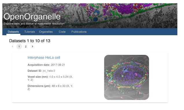
The resulting wealth of data arising from the task performed here, is unique and comprehensive, and covers multiple cells, with all their fine ultrastructural details at sub-organelle level. The authors made available 10 datasets containing 7 common wild-type cultured cell samples, and 3 different tissue samples. The authors made all the data available at https://openorganelle.janelia.org, which allows the wide scientific community to browse, download and explore the data. Within this database, the metadata and pre-selected views of interest are provided, while data can be further explored using an in-built visualization tool. Moreover, the repository contains segmentations into various categories of interest allowing quantification of numbers, sizes, shapes, contacts, locations, proximity, and multiple other parameters regarding organelles and proteins within the whole cell.
Key findings
- HeLa cell: HeLa cells are human cervical cancer cells widely used across multiple research fields. Having such cells imaged fully and to great detail, provides a strong reference point for the multiple conditions and studies for which these cells are models. The authors present here a whole HeLa cell including all its organelles, such as the mitochondrial network, the centrosome, the Golgi apparatus, and the nuclear envelope. The dataset already shows the advantages of 3D isotropic imaging, which provides details that no single 2D section would allow visualizing.
- Cytotoxic T cell attacking an ovarian cancer cell: The authors give this as an example of cell-cell interactions, focusing on the immunological synapse. They discuss that although the immunological synapse has been a main focus of interest, two-dimensional studies lack sufficient detail of this important interaction. Imaging of this interaction with FIB-SEM allowed a complete map of the complex membrane topology at the interface between the T cell and its target. Zooming into images allowed visualizing key features including membrane interdigitation, flat membrane apposition, and filopodia of the target cell trapped between two cells. Altogether, FIB-SEM allowed label-free localization of all organelles in the T cell, including those necessary for target killing and cytokine secretion.
- Mouse pancreatic islets. Pancreatic islets are microorgans consisting mainly of beta, alpha, delta polypeptide cells and endothelial cells. Among them, beta cells secrete insulin stored in secretory granules to maintain blood glucose homeostasis. Beta cells have a polyhedral shape, and whole cell imaging at high resolution is known to be challenging. Large-scale high-resolution FIB-SEM enables the analysis of ultrastructural differences between beta cells within an islet, as well as sub-cellular features, such as ribosomes or the cytoskeleton. In their work, the authors were able to image the cytoskeleton, as well as interactions between beta cells including primary cilia, their connections to neighbouring cells, and the intermingling of microvilli. They also report close contacts of the ER and insulin secretory granules in 3D, and provide a comprehensive 3D representation of microtubule networks and how they interact with other organelles. Moreover, they observed insulin secretory granules enriched near the plasma in association with microtubules, and independent of extracellular glucose levels.
- Drosophila brains. The authors investigated the central complex, and whether it contains characteristic synaptic motifs (including polyadic synapses, rosette synapses, or any new synaptic motif). They report that FIB-SEM allows reconstruction not only of large neuronal objects, but also smaller intracellular components such as synaptic vesicles, dense-core vesicles and microtubules.
What I like about this preprint
This is altogether a fascinating step forward both in the technology that enables this type of imaging, and in the fact that the resulting data was made publicly available to the scientific community and it can be further interacted with and used according to user-specific needs. I think reference databases in microscopy are often missing, and one such as this, with such impressive ultrastructural detail will be extremely valuable, and could set an important precedent for cell biology and imaging.
Open questions
- How accessible and/or easily implementable are the modifications you report for FIB-SEM? Do you envisage that many labs world-wide will be able to perform this imaging, and contribute to the fascinating database you have begun?
- You mention that the data you have acquired and which you discuss in this work is made available in https://openorganelle.janelia.org. Can groups that are able to perform eventually, this type of imaging and use the datasets you provide here as reference, upload their findings and comparisons to eg. pathological conditions of other sorts? What you have provided here is a fascinating basis for the scientific community.
- The time required to perform this detailed imaging is still very large. Many labs have the limitation that if only one microscope is available, it cannot be blocked for months at a time. Do you envisage that isotropic 3D EM will eventually be possible, albeit in shorter amounts of time?
- You tested your developments in various types of cells, and you discuss some of the obstacles presented by some of them. Do you envisage other limitations that readers and potential users should be aware of when exploring multiple types of samples?
References
- Xu et al, Isotropic 3D electron microscopy reference library of whole cells and tissues, bioRxiv, 2020.
doi: https://doi.org/10.1242/prelights.26566
Read preprint (No Ratings Yet)
Sign up to customise the site to your preferences and to receive alerts
Register hereAlso in the cell biology category:
Combinatorial and Inducible CRISPRa/i Enables Canalized hiPSC Forward Programming and Iterative Refinement via Single-Cell Genomics
Cell-ID
Developmental conversion of the nucleolus into an RNA Polymerase II transcriptional platform in Drosophila spermatocytes
Panagiotis Giannios
Cell position is more important than cell shape or age for the acquisition of cell identity in the brown alga Ectocarpus
Urvashi Goswami
preLists in the cell biology category:
Developmental regulation: molecular and ecological niches
This conference was held at the Station Biologique de Roscoff (France) and brought together researchers exploring how diverse niche environments shape developmental processes across scales. Spanning topics from ecological and metabolic influences to signalling networks, mechanics and gene regulation, the meeting highlighted the interplay between intrinsic and extrinsic factors in controlling cell fate and tissue organisation. This preList gathers preprints discussed by speakers and poster presenters during the meeting. Please do get in touch at preLights@biologists.com if you notice any relevant preprints that we may have missed.
| List by | Ingrid Tsang |
preLighters’ choice – Handpicked DevBio preprints
preLighters with expertise across developmental and stem cell biology have nominated a few developmental biology (and related) preprints they’re excited about and explain in a few paragraph why. Concise preprint highlights, prepared by the preLighter community – a quick way to spot upcoming trends, new methods and fresh ideas.
| List by | Theodora Stougiannou et al. |
BSDB Spring Meeting: Molecules to Morphogenesis
The British Society for Developmental Biology (BSDB) Spring Meeting Molecules to Morphogenesis was held from 23–26 March 2026 at the University of Warwick (UK). This meeting brought together a vibrant community of researchers to discuss how molecular mechanisms are integrated across scales to drive morphogenesis, spanning diverse model systems and approaches. This preList contains preprints by presenters from the talk and poster sessions at the meeting. Please do get in touch at preLights@biologists.com if you notice any relevant preprints that we may have missed.
| List by | Ingrid Tsang |
Keystone Symposium on Stem Cell Models in Embryology 2026
The Keystone Symposium on Stem Cell Models in Embryology, 2026, was organised by Jun Wu (UT Southwestern), Jianping Fu (University of Michigan) and Miki Ebisuya (TU Dresden) and held at Asilomar Conference Grounds in California (US). The meeting discussed recent advances made in establishing stem-cell-based embryo models, what fundamental insights into developmental processes have been gleaned from them, as well as how they are beginning to be applied more widely. This prelist contains preprints by presenters at the talk and poster sessions at the conference, which our Reviews Editor in attendance spotted. Please do reach out to preLights@biologists.com if you notice any that we’ve missed.
| List by | Ingrid Tsang |
SciELO preprints – From 2025 onwards
SciELO has become a cornerstone of open, multilingual scholarly communication across Latin America. Its preprint server, SciELO preprints, is expanding the global reach of preprinted research from the region (for more information, see our interview with Carolina Tanigushi). This preList brings together biological, English language SciELO preprints to help readers discover emerging work from the Global South. By highlighting these preprints in one place, we aim to support visibility, encourage early feedback, and showcase the vibrant research communities contributing to SciELO’s open science ecosystem.
| List by | Carolina Tanigushi |
November in preprints – DevBio & Stem cell biology
preLighters with expertise across developmental and stem cell biology have nominated a few developmental and stem cell biology (and related) preprints posted in November they’re excited about and explain in a single paragraph why. Concise preprint highlights, prepared by the preLighter community – a quick way to spot upcoming trends, new methods and fresh ideas.
| List by | Aline Grata et al. |
October in preprints – DevBio & Stem cell biology
Each month, preLighters with expertise across developmental and stem cell biology nominate a few recent developmental and stem cell biology (and related) preprints they’re excited about and explain in a single paragraph why. Short, snappy picks from working scientists — a quick way to spot fresh ideas, bold methods and papers worth reading in full. These preprints can all be found in the October preprint list published on the Node.
| List by | Deevitha Balasubramanian et al. |
October in preprints – Cell biology edition
Different preLighters, with expertise across cell biology, have worked together to create this preprint reading list for researchers with an interest in cell biology. This month, most picks fall under (1) Cell organelles and organisation, followed by (2) Mechanosignaling and mechanotransduction, (3) Cell cycle and division and (4) Cell migration
| List by | Matthew Davies et al. |
September in preprints – Cell biology edition
A group of preLighters, with expertise in different areas of cell biology, have worked together to create this preprint reading list. This month, categories include: (1) Cell organelles and organisation, (2) Cell signalling and mechanosensing, (3) Cell metabolism, (4) Cell cycle and division, (5) Cell migration
| List by | Sristilekha Nath et al. |
July in preprints – the CellBio edition
A group of preLighters, with expertise in different areas of cell biology, have worked together to create this preprint reading lists for researchers with an interest in cell biology. This month, categories include: (1) Cell Signalling and Mechanosensing (2) Cell Cycle and Division (3) Cell Migration and Cytoskeleton (4) Cancer Biology (5) Cell Organelles and Organisation
| List by | Girish Kale et al. |
June in preprints – the CellBio edition
A group of preLighters, with expertise in different areas of cell biology, have worked together to create this preprint reading lists for researchers with an interest in cell biology. This month, categories include: (1) Cell organelles and organisation (2) Cell signaling and mechanosensation (3) Genetics/gene expression (4) Biochemistry (5) Cytoskeleton
| List by | Barbora Knotkova et al. |
May in preprints – the CellBio edition
A group of preLighters, with expertise in different areas of cell biology, have worked together to create this preprint reading lists for researchers with an interest in cell biology. This month, categories include: 1) Biochemistry/metabolism 2) Cancer cell Biology 3) Cell adhesion, migration and cytoskeleton 4) Cell organelles and organisation 5) Cell signalling and 6) Genetics
| List by | Barbora Knotkova et al. |
Keystone Symposium – Metabolic and Nutritional Control of Development and Cell Fate
This preList contains preprints discussed during the Metabolic and Nutritional Control of Development and Cell Fate Keystone Symposia. This conference was organized by Lydia Finley and Ralph J. DeBerardinis and held in the Wylie Center and Tupper Manor at Endicott College, Beverly, MA, United States from May 7th to 9th 2025. This meeting marked the first in-person gathering of leading researchers exploring how metabolism influences development, including processes like cell fate, tissue patterning, and organ function, through nutrient availability and metabolic regulation. By integrating modern metabolic tools with genetic and epidemiological insights across model organisms, this event highlighted key mechanisms and identified open questions to advance the emerging field of developmental metabolism.
| List by | Virginia Savy, Martin Estermann |
April in preprints – the CellBio edition
A group of preLighters, with expertise in different areas of cell biology, have worked together to create this preprint reading lists for researchers with an interest in cell biology. This month, categories include: 1) biochemistry/metabolism 2) cell cycle and division 3) cell organelles and organisation 4) cell signalling and mechanosensing 5) (epi)genetics
| List by | Vibha SINGH et al. |
March in preprints – the CellBio edition
A group of preLighters, with expertise in different areas of cell biology, have worked together to create this preprint reading lists for researchers with an interest in cell biology. This month, categories include: 1) cancer biology 2) cell migration 3) cell organelles and organisation 4) cell signalling and mechanosensing 5) genetics and genomics 6) other
| List by | Girish Kale et al. |
Biologists @ 100 conference preList
This preList aims to capture all preprints being discussed at the Biologists @100 conference in Liverpool, UK, either as part of the poster sessions or the (flash/short/full-length) talks.
| List by | Reinier Prosee, Jonathan Townson |
February in preprints – the CellBio edition
A group of preLighters, with expertise in different areas of cell biology, have worked together to create this preprint reading lists for researchers with an interest in cell biology. This month, categories include: 1) biochemistry and cell metabolism 2) cell organelles and organisation 3) cell signalling, migration and mechanosensing
| List by | Barbora Knotkova et al. |
Community-driven preList – Immunology
In this community-driven preList, a group of preLighters, with expertise in different areas of immunology have worked together to create this preprint reading list.
| List by | Felipe Del Valle Batalla et al. |
January in preprints – the CellBio edition
A group of preLighters, with expertise in different areas of cell biology, have worked together to create this preprint reading lists for researchers with an interest in cell biology. This month, categories include: 1) biochemistry/metabolism 2) cell migration 3) cell organelles and organisation 4) cell signalling and mechanosensing 5) genetics/gene expression
| List by | Barbora Knotkova et al. |
December in preprints – the CellBio edition
A group of preLighters, with expertise in different areas of cell biology, have worked together to create this preprint reading lists for researchers with an interest in cell biology. This month, categories include: 1) cell cycle and division 2) cell migration and cytoskeleton 3) cell organelles and organisation 4) cell signalling and mechanosensing 5) genetics/gene expression
| List by | Matthew Davies et al. |
November in preprints – the CellBio edition
This is the first community-driven preList! A group of preLighters, with expertise in different areas of cell biology, have worked together to create this preprint reading lists for researchers with an interest in cell biology. Categories include: 1) cancer cell biology 2) cell cycle and division 3) cell migration and cytoskeleton 4) cell organelles and organisation 5) cell signalling and mechanosensing 6) genetics/gene expression
| List by | Felipe Del Valle Batalla et al. |
BSCB-Biochemical Society 2024 Cell Migration meeting
This preList features preprints that were discussed and presented during the BSCB-Biochemical Society 2024 Cell Migration meeting in Birmingham, UK in April 2024. Kindly put together by Sara Morais da Silva, Reviews Editor at Journal of Cell Science.
| List by | Reinier Prosee |
‘In preprints’ from Development 2022-2023
A list of the preprints featured in Development's 'In preprints' articles between 2022-2023
| List by | Alex Eve, Katherine Brown |
preLights peer support – preprints of interest
This is a preprint repository to organise the preprints and preLights covered through the 'preLights peer support' initiative.
| List by | preLights peer support |
The Society for Developmental Biology 82nd Annual Meeting
This preList is made up of the preprints discussed during the Society for Developmental Biology 82nd Annual Meeting that took place in Chicago in July 2023.
| List by | Joyce Yu, Katherine Brown |
CSHL 87th Symposium: Stem Cells
Preprints mentioned by speakers at the #CSHLsymp23
| List by | Alex Eve |
Journal of Cell Science meeting ‘Imaging Cell Dynamics’
This preList highlights the preprints discussed at the JCS meeting 'Imaging Cell Dynamics'. The meeting was held from 14 - 17 May 2023 in Lisbon, Portugal and was organised by Erika Holzbaur, Jennifer Lippincott-Schwartz, Rob Parton and Michael Way.
| List by | Helen Zenner |
9th International Symposium on the Biology of Vertebrate Sex Determination
This preList contains preprints discussed during the 9th International Symposium on the Biology of Vertebrate Sex Determination. This conference was held in Kona, Hawaii from April 17th to 21st 2023.
| List by | Martin Estermann |
Alumni picks – preLights 5th Birthday
This preList contains preprints that were picked and highlighted by preLights Alumni - an initiative that was set up to mark preLights 5th birthday. More entries will follow throughout February and March 2023.
| List by | Sergio Menchero et al. |
CellBio 2022 – An ASCB/EMBO Meeting
This preLists features preprints that were discussed and presented during the CellBio 2022 meeting in Washington, DC in December 2022.
| List by | Nadja Hümpfer et al. |
Fibroblasts
The advances in fibroblast biology preList explores the recent discoveries and preprints of the fibroblast world. Get ready to immerse yourself with this list created for fibroblasts aficionados and lovers, and beyond. Here, my goal is to include preprints of fibroblast biology, heterogeneity, fate, extracellular matrix, behavior, topography, single-cell atlases, spatial transcriptomics, and their matrix!
| List by | Osvaldo Contreras |
EMBL Synthetic Morphogenesis: From Gene Circuits to Tissue Architecture (2021)
A list of preprints mentioned at the #EESmorphoG virtual meeting in 2021.
| List by | Alex Eve |
FENS 2020
A collection of preprints presented during the virtual meeting of the Federation of European Neuroscience Societies (FENS) in 2020
| List by | Ana Dorrego-Rivas |
Planar Cell Polarity – PCP
This preList contains preprints about the latest findings on Planar Cell Polarity (PCP) in various model organisms at the molecular, cellular and tissue levels.
| List by | Ana Dorrego-Rivas |
BioMalPar XVI: Biology and Pathology of the Malaria Parasite
[under construction] Preprints presented at the (fully virtual) EMBL BioMalPar XVI, 17-18 May 2020 #emblmalaria
| List by | Dey Lab, Samantha Seah |
1
Cell Polarity
Recent research from the field of cell polarity is summarized in this list of preprints. It comprises of studies focusing on various forms of cell polarity ranging from epithelial polarity, planar cell polarity to front-to-rear polarity.
| List by | Yamini Ravichandran |
TAGC 2020
Preprints recently presented at the virtual Allied Genetics Conference, April 22-26, 2020. #TAGC20
| List by | Maiko Kitaoka et al. |
3D Gastruloids
A curated list of preprints related to Gastruloids (in vitro models of early development obtained by 3D aggregation of embryonic cells). Updated until July 2021.
| List by | Paul Gerald L. Sanchez and Stefano Vianello |
ECFG15 – Fungal biology
Preprints presented at 15th European Conference on Fungal Genetics 17-20 February 2020 Rome
| List by | Hiral Shah |
ASCB EMBO Annual Meeting 2019
A collection of preprints presented at the 2019 ASCB EMBO Meeting in Washington, DC (December 7-11)
| List by | Madhuja Samaddar et al. |
EMBL Seeing is Believing – Imaging the Molecular Processes of Life
Preprints discussed at the 2019 edition of Seeing is Believing, at EMBL Heidelberg from the 9th-12th October 2019
| List by | Dey Lab |
Autophagy
Preprints on autophagy and lysosomal degradation and its role in neurodegeneration and disease. Includes molecular mechanisms, upstream signalling and regulation as well as studies on pharmaceutical interventions to upregulate the process.
| List by | Sandra Malmgren Hill |
Lung Disease and Regeneration
This preprint list compiles highlights from the field of lung biology.
| List by | Rob Hynds |
Cellular metabolism
A curated list of preprints related to cellular metabolism at Biorxiv by Pablo Ranea Robles from the Prelights community. Special interest on lipid metabolism, peroxisomes and mitochondria.
| List by | Pablo Ranea Robles |
BSCB/BSDB Annual Meeting 2019
Preprints presented at the BSCB/BSDB Annual Meeting 2019
| List by | Dey Lab |
MitoList
This list of preprints is focused on work expanding our knowledge on mitochondria in any organism, tissue or cell type, from the normal biology to the pathology.
| List by | Sandra Franco Iborra |
Biophysical Society Annual Meeting 2019
Few of the preprints that were discussed in the recent BPS annual meeting at Baltimore, USA
| List by | Joseph Jose Thottacherry |
ASCB/EMBO Annual Meeting 2018
This list relates to preprints that were discussed at the recent ASCB conference.
| List by | Dey Lab, Amanda Haage |