


Membrane Curvature Promotes ER-PM Contact Formation via Junctophilin-EHD Interactions
Posted on: 7 August 2024 , updated on: 8 August 2024
Preprint posted on 1 July 2024
This is how membrane curvature makes our hearts beat - Junctophilins interact with EHD proteins to connect the sarcoplasm to t-tubules and thus enable excitation-contraction coupling in heart muscle cells.
Selected by Barbora KnotkovaCategories: cell biology
Background:
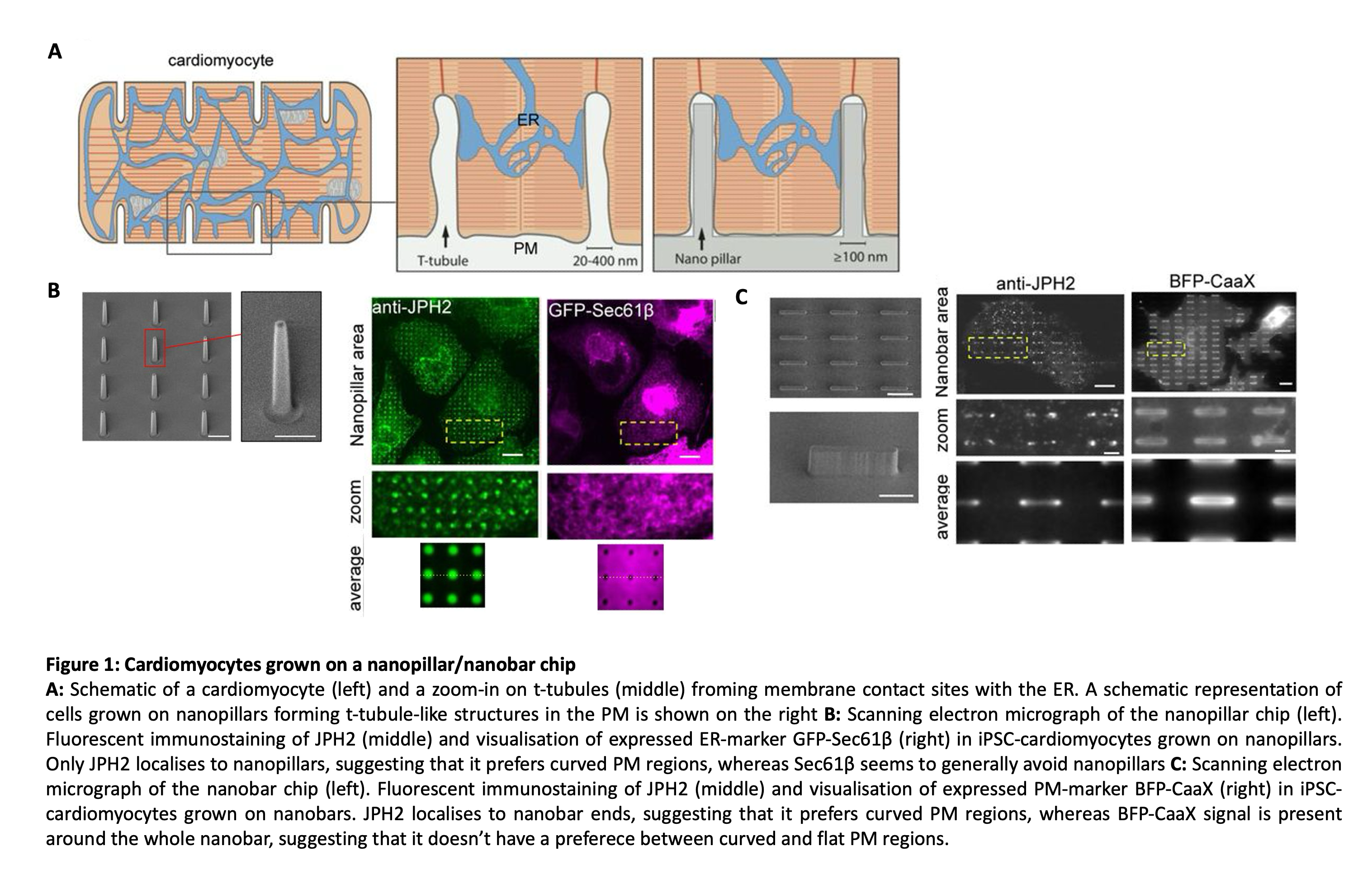
Cardiomyocytes receive signals to contract in the form of action potentials at long invaginations of the plasma membrane (PM) called transverse tubules (t-tubules). T-tubules form extensive membrane contact sites with the endoplasmic reticulum (ER) called dyad junctions (see Figure 1A). Dyad junctions are important for passing the signal on to sarcomeres, the contractile units of cardiomyocytes: When the PM depolarises upon action potential arrival, voltage-gated L-type calcium channels (LTCCs) in the t-tubules open and calcium ions enter the cytosolic gap between the t-tubule and the ER. This influx of calcium in turn leads to the opening of ryanodine receptor (RyR) channels in the ER, which release even higher quantities of calcium into the cytoplasm. Calcium then binds to the protein troponinin in the sarcomere and enables myosin to interact with actin filaments, leading to muscle contraction. The co-localisation of the PM-localised LTCC channels and the ER-localised RyR channels is clearly important for efficient calcium signalling and muscle contraction. The proteins responsible for co-localisation of the channels at dyad junctions are called junctophilins (JPHs). However, how junctophilins themselves are sorted to the dyad junction has until now been unknown.
In this preprint, Yang and colleagues decipher how junctophilins localise to curved plasma membrane regions. They show that Epsin15-homology domain containing proteins (EHDs) are required for junctophilin recruitment to curved membranes and identify the junctophilin domains required for EHD binding.
Method:
Different cell types, including cardiomyocytes that were either induced pluripotent stem cell differentiated (iPSC) or taken from rat embryos and the osteosarcoma cell line U2OS, were cultured on nanochips dotted with a regular array of nanopillar or nanobar structures. The cells wrap around these structures leading to curvature of the plasma membrane. Nanopillars with a diameter of 200-300 nm were used to resemble t-tubules. In the case of nanobars, a width of 200 nm and a length of 2 µm was used, leading to areas of curved and straight plasma membrane. Using fluorescent microscopy, the localisation of proteins could be investigated with regard to the nanopillar vs the flat surface in between, and with regard to the curved ends vs the flat sides of the nanobar (see Figure 1).
Key findings:
-
- Components of dyad junctions localise to curved membranes in cells grown on nanopillar or nanobar grids
-
-
- The authors first checked the distribution of immuno-stained junctophilin JPH2 in iPSC-cardiomyocytes, which lack t-tubules, grown on the curvature-inducing surface. They observed that JPH2 accumulated at nanopillars or the ends of a nanobar, but the ER-resident protein translocon Sec61 was dispersed.
- Other components of the dyad junction, such as LTCC (calcium channel in the PM) and RyR2 (calcium channel in the ER) also localised to the highly-curved membrane regions while the PM marker BFP-CAAX was present around the whole nanopillar.
- E-Syt2 (another ER-PM tether), on the other hand, localised to the side walls (=flat membrane) of nanobars, showing that curvature preference is specific to junctophilin-mediated ER-PM contact sites.
- Focused ion beam scanning electron microscopy revealed that ER-PM contacts preferentially form at nanopillars in cardiomyocytes. In WT U2OS cells, the ER-PM contact site marker MAPPER localised to nanobar ends. Both findings suggest that ER-PM contacts preferentially form at curved PM.
-
- STIM1 and ORAI1 are recruited to ER-PM contact sites at curved regions of the PM upon ER calcium store depletion
-
-
- Before calcium store depletion, STIM1 was found in the intracellular ER network and ORAI1 was diffused throughout the PM.
- After thapsigargin-induced ER calcium store depletion, STIM1 and ORAI1 co-localised at nanobar ends, suggesting that the proteins are preferentially recruited to ER-PM contact sites at curved PM.
-
- The low complexity region (LCR) and the MORN repeats domain of JPH3 are both required for targeting to curved PM
-
-
- The MORN domain as well as LCR could bind to the PM independently of each other – deletion of just one did not hinder PM binding and when expressed in isolation, each localised to the PM. However, all of these contructs lost preference for curvature, indicating that both, the MORN domain and the LCR, are required for binding to curved PM regions.
- The authors furthermore identified a polybasic motif within the LCR, which is required for PM binding, and showed that mutations of serine residues in the MORN/LCR region led to loss of curvature localisation. This is very interesting from a clinical point of view as these mutations are found in patients with hypertrophic cardiomyopathy.
-
- JPHs bind to EHD proteins which are known to be important for t-tubule morphology
-
-
- Out of all JPH2-interacting proteins that are involved in curvature-dependent processes at the PM, only triple knock-down of EHD1, 2 and 4 disrupted JPH3’s localisation to nanobar ends.
- Extraction of cholesterol changes the localisation of EHDs. Here the authors could show that this treatment disrupts not only EHD4’s localisation to nanobar ends but also that of JPH3.
- Both, the MORN repeats and the LCR region were needed for binding to EHD as shown by co-localisation of MORN-LCR constructs with EHD4 in cells grown on a flat surface. Furthermore, only a construct containing both domains could be co-immunoprecipitated with EHD4.
-
What I like about the preprint:
This preprint initially caught my interest because it studies a membrane contact site that preferentially forms at a curved membrane. This architectural arrangement strikingly resembles the membrane contact site between the inner and outer mitochondrial membrane, which I investigate in my PhD research. I also really like the method of artificially inducing curvature in live cells with nanostructures. It is something I have not come across before and it has yielded really fascinating fluorescent images in this study.
Questions for the authors:
- As far as I understood, JPH proteins are predominantly expressed in muscles and neurons. Why then are JPH3 and 4 expressed in osteosarcoma U2OS cells? Could they, together with EHDs, facilitate ER-PM membrane contact sites with other functions? Are there any known curved PM regions in U2OS cells that form ER contacts or may your findings lead to the discovery of new processes requiring ER-PM membrane contact sites at curved PM?
- Can LCR with mutated polybasic region still bind to EHDs? Can pathogenic mutant variants still bind to EHDs?
- Could you quantify the extent of ER-PM contact sites at nanopillars in WT cells vs cells with disrupted EHD-Junctophilin binding (or cells with cholesterol extracted EHD / EHD or junctophilin KD)? It would be interesting to know whether the junctophilin tether is required to localise PM-ER contact sites to the nanopillar or whether there are other tethers with curvature preference. In connection to this, does loss of EHD-junctophilin interaction lead to loss of LTCC and RyR2 colocalisation at nanopillars?
Bibliography:
Bhattacharyya, S. and T. J. Pucadyil (2020). “Cellular functions and intrinsic attributes of the ATP-binding Eps15 homology domain-containing proteins.” Protein Sci 29(6): 1321-1330. DOI: 10.1002/pro.3860
Lu, F. and W. T. Pu (2020). “The architecture and function of cardiac dyads.” Biophys Rev 12(4): 1007-1017. DOI: 10.1007/s12551-020-00729-x
Piggott, C. A. and Y. Jin (2021). “Junctophilins: Key Membrane Tethers in Muscles and Neurons.” Front Mol Neurosci 14: 709390. DOI: 10.3389/fnmol.2021.709390. DOI: 10.3389/fnmol.2021.709390
doi: https://doi.org/10.1242/prelights.38028
Read preprint (No Ratings Yet)
Sign up to customise the site to your preferences and to receive alerts
Register hereAlso in the cell biology category:
Combinatorial and Inducible CRISPRa/i Enables Canalized hiPSC Forward Programming and Iterative Refinement via Single-Cell Genomics
Cell-ID
Developmental conversion of the nucleolus into an RNA Polymerase II transcriptional platform in Drosophila spermatocytes
Panagiotis Giannios
Cell position is more important than cell shape or age for the acquisition of cell identity in the brown alga Ectocarpus
Urvashi Goswami
preLists in the cell biology category:
Developmental regulation: molecular and ecological niches
This conference was held at the Station Biologique de Roscoff (France) and brought together researchers exploring how diverse niche environments shape developmental processes across scales. Spanning topics from ecological and metabolic influences to signalling networks, mechanics and gene regulation, the meeting highlighted the interplay between intrinsic and extrinsic factors in controlling cell fate and tissue organisation. This preList gathers preprints discussed by speakers and poster presenters during the meeting. Please do get in touch at preLights@biologists.com if you notice any relevant preprints that we may have missed.
| List by | Ingrid Tsang |
preLighters’ choice – Handpicked DevBio preprints
preLighters with expertise across developmental and stem cell biology have nominated a few developmental biology (and related) preprints they’re excited about and explain in a few paragraph why. Concise preprint highlights, prepared by the preLighter community – a quick way to spot upcoming trends, new methods and fresh ideas.
| List by | Theodora Stougiannou et al. |
BSDB Spring Meeting: Molecules to Morphogenesis
The British Society for Developmental Biology (BSDB) Spring Meeting Molecules to Morphogenesis was held from 23–26 March 2026 at the University of Warwick (UK). This meeting brought together a vibrant community of researchers to discuss how molecular mechanisms are integrated across scales to drive morphogenesis, spanning diverse model systems and approaches. This preList contains preprints by presenters from the talk and poster sessions at the meeting. Please do get in touch at preLights@biologists.com if you notice any relevant preprints that we may have missed.
| List by | Ingrid Tsang |
Keystone Symposium on Stem Cell Models in Embryology 2026
The Keystone Symposium on Stem Cell Models in Embryology, 2026, was organised by Jun Wu (UT Southwestern), Jianping Fu (University of Michigan) and Miki Ebisuya (TU Dresden) and held at Asilomar Conference Grounds in California (US). The meeting discussed recent advances made in establishing stem-cell-based embryo models, what fundamental insights into developmental processes have been gleaned from them, as well as how they are beginning to be applied more widely. This prelist contains preprints by presenters at the talk and poster sessions at the conference, which our Reviews Editor in attendance spotted. Please do reach out to preLights@biologists.com if you notice any that we’ve missed.
| List by | Ingrid Tsang |
SciELO preprints – From 2025 onwards
SciELO has become a cornerstone of open, multilingual scholarly communication across Latin America. Its preprint server, SciELO preprints, is expanding the global reach of preprinted research from the region (for more information, see our interview with Carolina Tanigushi). This preList brings together biological, English language SciELO preprints to help readers discover emerging work from the Global South. By highlighting these preprints in one place, we aim to support visibility, encourage early feedback, and showcase the vibrant research communities contributing to SciELO’s open science ecosystem.
| List by | Carolina Tanigushi |
November in preprints – DevBio & Stem cell biology
preLighters with expertise across developmental and stem cell biology have nominated a few developmental and stem cell biology (and related) preprints posted in November they’re excited about and explain in a single paragraph why. Concise preprint highlights, prepared by the preLighter community – a quick way to spot upcoming trends, new methods and fresh ideas.
| List by | Aline Grata et al. |
October in preprints – DevBio & Stem cell biology
Each month, preLighters with expertise across developmental and stem cell biology nominate a few recent developmental and stem cell biology (and related) preprints they’re excited about and explain in a single paragraph why. Short, snappy picks from working scientists — a quick way to spot fresh ideas, bold methods and papers worth reading in full. These preprints can all be found in the October preprint list published on the Node.
| List by | Deevitha Balasubramanian et al. |
October in preprints – Cell biology edition
Different preLighters, with expertise across cell biology, have worked together to create this preprint reading list for researchers with an interest in cell biology. This month, most picks fall under (1) Cell organelles and organisation, followed by (2) Mechanosignaling and mechanotransduction, (3) Cell cycle and division and (4) Cell migration
| List by | Matthew Davies et al. |
September in preprints – Cell biology edition
A group of preLighters, with expertise in different areas of cell biology, have worked together to create this preprint reading list. This month, categories include: (1) Cell organelles and organisation, (2) Cell signalling and mechanosensing, (3) Cell metabolism, (4) Cell cycle and division, (5) Cell migration
| List by | Sristilekha Nath et al. |
July in preprints – the CellBio edition
A group of preLighters, with expertise in different areas of cell biology, have worked together to create this preprint reading lists for researchers with an interest in cell biology. This month, categories include: (1) Cell Signalling and Mechanosensing (2) Cell Cycle and Division (3) Cell Migration and Cytoskeleton (4) Cancer Biology (5) Cell Organelles and Organisation
| List by | Girish Kale et al. |
June in preprints – the CellBio edition
A group of preLighters, with expertise in different areas of cell biology, have worked together to create this preprint reading lists for researchers with an interest in cell biology. This month, categories include: (1) Cell organelles and organisation (2) Cell signaling and mechanosensation (3) Genetics/gene expression (4) Biochemistry (5) Cytoskeleton
| List by | Barbora Knotkova et al. |
May in preprints – the CellBio edition
A group of preLighters, with expertise in different areas of cell biology, have worked together to create this preprint reading lists for researchers with an interest in cell biology. This month, categories include: 1) Biochemistry/metabolism 2) Cancer cell Biology 3) Cell adhesion, migration and cytoskeleton 4) Cell organelles and organisation 5) Cell signalling and 6) Genetics
| List by | Barbora Knotkova et al. |
Keystone Symposium – Metabolic and Nutritional Control of Development and Cell Fate
This preList contains preprints discussed during the Metabolic and Nutritional Control of Development and Cell Fate Keystone Symposia. This conference was organized by Lydia Finley and Ralph J. DeBerardinis and held in the Wylie Center and Tupper Manor at Endicott College, Beverly, MA, United States from May 7th to 9th 2025. This meeting marked the first in-person gathering of leading researchers exploring how metabolism influences development, including processes like cell fate, tissue patterning, and organ function, through nutrient availability and metabolic regulation. By integrating modern metabolic tools with genetic and epidemiological insights across model organisms, this event highlighted key mechanisms and identified open questions to advance the emerging field of developmental metabolism.
| List by | Virginia Savy, Martin Estermann |
April in preprints – the CellBio edition
A group of preLighters, with expertise in different areas of cell biology, have worked together to create this preprint reading lists for researchers with an interest in cell biology. This month, categories include: 1) biochemistry/metabolism 2) cell cycle and division 3) cell organelles and organisation 4) cell signalling and mechanosensing 5) (epi)genetics
| List by | Vibha SINGH et al. |
March in preprints – the CellBio edition
A group of preLighters, with expertise in different areas of cell biology, have worked together to create this preprint reading lists for researchers with an interest in cell biology. This month, categories include: 1) cancer biology 2) cell migration 3) cell organelles and organisation 4) cell signalling and mechanosensing 5) genetics and genomics 6) other
| List by | Girish Kale et al. |
Biologists @ 100 conference preList
This preList aims to capture all preprints being discussed at the Biologists @100 conference in Liverpool, UK, either as part of the poster sessions or the (flash/short/full-length) talks.
| List by | Reinier Prosee, Jonathan Townson |
February in preprints – the CellBio edition
A group of preLighters, with expertise in different areas of cell biology, have worked together to create this preprint reading lists for researchers with an interest in cell biology. This month, categories include: 1) biochemistry and cell metabolism 2) cell organelles and organisation 3) cell signalling, migration and mechanosensing
| List by | Barbora Knotkova et al. |
Community-driven preList – Immunology
In this community-driven preList, a group of preLighters, with expertise in different areas of immunology have worked together to create this preprint reading list.
| List by | Felipe Del Valle Batalla et al. |
January in preprints – the CellBio edition
A group of preLighters, with expertise in different areas of cell biology, have worked together to create this preprint reading lists for researchers with an interest in cell biology. This month, categories include: 1) biochemistry/metabolism 2) cell migration 3) cell organelles and organisation 4) cell signalling and mechanosensing 5) genetics/gene expression
| List by | Barbora Knotkova et al. |
December in preprints – the CellBio edition
A group of preLighters, with expertise in different areas of cell biology, have worked together to create this preprint reading lists for researchers with an interest in cell biology. This month, categories include: 1) cell cycle and division 2) cell migration and cytoskeleton 3) cell organelles and organisation 4) cell signalling and mechanosensing 5) genetics/gene expression
| List by | Matthew Davies et al. |
November in preprints – the CellBio edition
This is the first community-driven preList! A group of preLighters, with expertise in different areas of cell biology, have worked together to create this preprint reading lists for researchers with an interest in cell biology. Categories include: 1) cancer cell biology 2) cell cycle and division 3) cell migration and cytoskeleton 4) cell organelles and organisation 5) cell signalling and mechanosensing 6) genetics/gene expression
| List by | Felipe Del Valle Batalla et al. |
BSCB-Biochemical Society 2024 Cell Migration meeting
This preList features preprints that were discussed and presented during the BSCB-Biochemical Society 2024 Cell Migration meeting in Birmingham, UK in April 2024. Kindly put together by Sara Morais da Silva, Reviews Editor at Journal of Cell Science.
| List by | Reinier Prosee |
‘In preprints’ from Development 2022-2023
A list of the preprints featured in Development's 'In preprints' articles between 2022-2023
| List by | Alex Eve, Katherine Brown |
preLights peer support – preprints of interest
This is a preprint repository to organise the preprints and preLights covered through the 'preLights peer support' initiative.
| List by | preLights peer support |
The Society for Developmental Biology 82nd Annual Meeting
This preList is made up of the preprints discussed during the Society for Developmental Biology 82nd Annual Meeting that took place in Chicago in July 2023.
| List by | Joyce Yu, Katherine Brown |
CSHL 87th Symposium: Stem Cells
Preprints mentioned by speakers at the #CSHLsymp23
| List by | Alex Eve |
Journal of Cell Science meeting ‘Imaging Cell Dynamics’
This preList highlights the preprints discussed at the JCS meeting 'Imaging Cell Dynamics'. The meeting was held from 14 - 17 May 2023 in Lisbon, Portugal and was organised by Erika Holzbaur, Jennifer Lippincott-Schwartz, Rob Parton and Michael Way.
| List by | Helen Zenner |
9th International Symposium on the Biology of Vertebrate Sex Determination
This preList contains preprints discussed during the 9th International Symposium on the Biology of Vertebrate Sex Determination. This conference was held in Kona, Hawaii from April 17th to 21st 2023.
| List by | Martin Estermann |
Alumni picks – preLights 5th Birthday
This preList contains preprints that were picked and highlighted by preLights Alumni - an initiative that was set up to mark preLights 5th birthday. More entries will follow throughout February and March 2023.
| List by | Sergio Menchero et al. |
CellBio 2022 – An ASCB/EMBO Meeting
This preLists features preprints that were discussed and presented during the CellBio 2022 meeting in Washington, DC in December 2022.
| List by | Nadja Hümpfer et al. |
Fibroblasts
The advances in fibroblast biology preList explores the recent discoveries and preprints of the fibroblast world. Get ready to immerse yourself with this list created for fibroblasts aficionados and lovers, and beyond. Here, my goal is to include preprints of fibroblast biology, heterogeneity, fate, extracellular matrix, behavior, topography, single-cell atlases, spatial transcriptomics, and their matrix!
| List by | Osvaldo Contreras |
EMBL Synthetic Morphogenesis: From Gene Circuits to Tissue Architecture (2021)
A list of preprints mentioned at the #EESmorphoG virtual meeting in 2021.
| List by | Alex Eve |
FENS 2020
A collection of preprints presented during the virtual meeting of the Federation of European Neuroscience Societies (FENS) in 2020
| List by | Ana Dorrego-Rivas |
Planar Cell Polarity – PCP
This preList contains preprints about the latest findings on Planar Cell Polarity (PCP) in various model organisms at the molecular, cellular and tissue levels.
| List by | Ana Dorrego-Rivas |
BioMalPar XVI: Biology and Pathology of the Malaria Parasite
[under construction] Preprints presented at the (fully virtual) EMBL BioMalPar XVI, 17-18 May 2020 #emblmalaria
| List by | Dey Lab, Samantha Seah |
1
Cell Polarity
Recent research from the field of cell polarity is summarized in this list of preprints. It comprises of studies focusing on various forms of cell polarity ranging from epithelial polarity, planar cell polarity to front-to-rear polarity.
| List by | Yamini Ravichandran |
TAGC 2020
Preprints recently presented at the virtual Allied Genetics Conference, April 22-26, 2020. #TAGC20
| List by | Maiko Kitaoka et al. |
3D Gastruloids
A curated list of preprints related to Gastruloids (in vitro models of early development obtained by 3D aggregation of embryonic cells). Updated until July 2021.
| List by | Paul Gerald L. Sanchez and Stefano Vianello |
ECFG15 – Fungal biology
Preprints presented at 15th European Conference on Fungal Genetics 17-20 February 2020 Rome
| List by | Hiral Shah |
ASCB EMBO Annual Meeting 2019
A collection of preprints presented at the 2019 ASCB EMBO Meeting in Washington, DC (December 7-11)
| List by | Madhuja Samaddar et al. |
EMBL Seeing is Believing – Imaging the Molecular Processes of Life
Preprints discussed at the 2019 edition of Seeing is Believing, at EMBL Heidelberg from the 9th-12th October 2019
| List by | Dey Lab |
Autophagy
Preprints on autophagy and lysosomal degradation and its role in neurodegeneration and disease. Includes molecular mechanisms, upstream signalling and regulation as well as studies on pharmaceutical interventions to upregulate the process.
| List by | Sandra Malmgren Hill |
Lung Disease and Regeneration
This preprint list compiles highlights from the field of lung biology.
| List by | Rob Hynds |
Cellular metabolism
A curated list of preprints related to cellular metabolism at Biorxiv by Pablo Ranea Robles from the Prelights community. Special interest on lipid metabolism, peroxisomes and mitochondria.
| List by | Pablo Ranea Robles |
BSCB/BSDB Annual Meeting 2019
Preprints presented at the BSCB/BSDB Annual Meeting 2019
| List by | Dey Lab |
MitoList
This list of preprints is focused on work expanding our knowledge on mitochondria in any organism, tissue or cell type, from the normal biology to the pathology.
| List by | Sandra Franco Iborra |
Biophysical Society Annual Meeting 2019
Few of the preprints that were discussed in the recent BPS annual meeting at Baltimore, USA
| List by | Joseph Jose Thottacherry |
ASCB/EMBO Annual Meeting 2018
This list relates to preprints that were discussed at the recent ASCB conference.
| List by | Dey Lab, Amanda Haage |