


Actomyosin forces and the energetics of red blood cell invasion by the malaria parasite Plasmodium falciparum
Posted on: 13 July 2020
Preprint posted on 25 June 2020
Article now published in PLOS Pathogens at http://dx.doi.org/10.1371/journal.ppat.1009007
Categories: cell biology
Background
Malaria is caused by single-celled, obligate intracellular parasites from the genus Plasmodium, the most virulent of which is Plasmodium falciparum. All symptoms associated with malaria are the result of the asexual erythrocytic stages, involving cycles of red blood cell (RBC) invasion and egress by the Plasmodium spp. merozoite. Merozoites are small cells around 1 µm in size, that use substrate-dependent gliding motility. Merozoites are primed for invasion by phosphorylation of the motility apparatus before RBC egress, and have a short window of viability to invade a RBC. Plasmodium merozoites rely on a conserved molecular motor for gliding and RBC invasion, centred around a MyoA motor complex (MMC) or glideosome, situated between the parasite plasma membrane and the double membrane inner membrane complex (IMC). However, the precise function of PfMyoA in invasion, its regulation, the role of other myosins and overall energetics of invasion remain unclear. Questions still remain open, for example, regarding PfMyoA organisation and function, how motor force is integrated with retrograde flow of parasite plasma membrane and how force is applied across the parasite. Altogether, a greater understanding of the mechanical function of actomyosin during merozoite invasion is important. In their work, Blake et al (1) combine a conditional mutagenesis strategy with live video microscopy to gain insight into the energetics of invasion and probe PfMyoA function, and that of the auxiliary motor PfMyoB, during merozoite invasion.
Key findings and developments
Parasite line generation and characterization
The authors began by generating an ectopic expression platform for Plasmodium myosins. They modified a previously existing line, a conditional KO for PfMyoA (PfMyoA-cKO) that excises a terminal portion of the PfMyoA gene on rapamycin (RAP) treatment rendering it non-functional (2), to further investigate the role of PfMyoA.
Altogether, they used a) the PfMyoA-cKO parasite line and generated de novo b) a line for PfMyoA-complementation (PfMyoA-comp); c) a line carrying a K764E mutation, which is designed to probe phospho-regulation of PfMyoA –(hypothetically, this mutation should leave merozoites unaffected if they only need PfMyoA for maximal force production during invasion); d) a mutant for the PfMyoA light chain ELC (PfELC-cKO), as previous studies have suggested that the absence of PfELC leaves a functional but strongly weakened motor; and e) a conditional KO for PfMyoB, as myosin redundancy has been shown to contribute to residual invasion in T. gondii MyoA-cKO parasites (3). All cKO phenotypes, like PfMyoA-cKO, could be initiated with RAP treatment (activating DiCre-dependent gene excision).
Time-lapse microscopy-based phenotype characterization
WT and DMSO-treated parasites
Time-lapse imaging in live cells has been consistently used to describe merozoite invasion and to characterize Plasmodium mutants at various stages. The authors went on to use live imaging to analyse merozoite invasion by the various mutant lines generated in this work. Parasites were synchronized in the schizont stage, and merozoite invasion characterized. Invasion phenotypes were characterized based on separate stages generally observed in WT parasites, namely, attachment to RBC, deformation of the RBC surface, internalization into the RBC, and RBC echinocytosis. Mutants were classified into 4 types: Type A: successful invasion; Type B: incomplete internalization and merozoite ejection; Type C: deformation present, but no internalization; and Type D: neither deformation not internalization present, just attachment. Moreover, a score was used to assign each event based on the intensity of deformation.
PfMyoA-cKO and PfMyoA-comp
PfMyoA-cKO parasites after RAP treatment showed zero successful invasion events, showing no deformation or internalization (Type D failure), and therefore this mutant could not be used to probe the role of the motor in internalization directly. For PfMyoA-comp parasites, whilst a moderate drop in invasion success was observed, no significant differences in deformation strength or phase timings were observed.
PfELC-cKO
Like PfMyoA-cKO parasites, rapamycin-treated PfELC-cKO parasites did not achieve any successful invasion. However, when observed in detail a significant proportion of events were nonetheless able to deform the RBC. This shows that a partially functional motor can achieve inefficient deformation. Furthermore, no PfELC-cKO merozoites were able to initiate internalization supporting a critical role for PfMyoA in driving merozoite internalisation, as well as deformation, and suggesting that the process of internalisation has a higher energetic barrier than deformation. Altogether, the results from PfMyoA-cKO and PfELC-cKO mutants shows that without PfMyoA or PfELC, merozoites cannot strongly deform or internalize.
PfMyoA-K764E
Understanding the role of motor force during internalisation depends on finding intermediate-strength motor mutants able to initiate internalisation (namely, a gradient effect). PfMyoA-K764E merozoites could initiate invasion, but showed a significant increase in Type B failures (being ejected from the RBC). PfMyoA-K764E parasites are more likely to fail at initiation of internalisation (an increase in Type C failures) and, when they can initiate it, they take longer to do so (a longer pre-internalisation pause). These parasites are also more likely to fail to complete internalisation (causing the increase in Type B failures) and they take much longer to internalise when they fail, and slightly longer even when successful.
PfMyoB-cKO
Video microscopy of RAP-treated PfMyoB-cKO parasites showed only a moderate reduction in successful invasion and increase in Type C failures, while the distribution of deformation scores was unchanged. The durations of some invasion phases were affected by PfMyoB-cKO. The significant delay in initiation of internalisation is consistent with a model of PfMyoB supporting the first stages of translocating the tight junction, but overall, PfMyoB is not required for internalization.
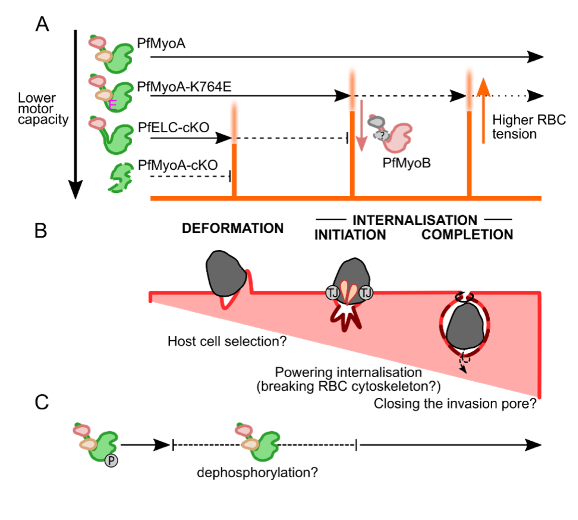
The study concludes overall, that while PfMyoB-cKO merozoites are delayed in initiation of internalisation, and PfMyoA-cKO and PfELC-cKO merozoites have insufficient force production to overcome the steps of deformation or internalisation, PfMyoA-K764E merozoites show a distinct defect at a third energetic barrier: completion of internalisation. These data therefore support a three-step model for the energetics of red blood cell entry by the merozoite: surface deformation of the RBC; initiation of internalisation/entry; and completion of internationalisation/closure of the tight junction around the entering merozoite.
What I like about this preprint
I find the question approached an interesting part of the puzzle to understand the invasion process. I like also that the authors took vast advantage of microscopy tools to explore the question addressed.
References
- Blake TCA et al, Actomyosin forces and the energetics of red blood cell invasion by the malaria parasite Plasmodium falciparum, bioRxiv, 2020.
- Birnbaum J et al, A genetic system to study Plasmodium falciparum protein function, Nature Methods, 2017.
- Frenal K et al, Plasticity between MyoC and MyoA- glideosomes: an example of functional compensation in Toxoplasma gondii PloS Pathogens, 2014.
doi: https://doi.org/10.1242/prelights.23049
Read preprint (No Ratings Yet)
Sign up to customise the site to your preferences and to receive alerts
Register hereAlso in the cell biology category:
Combinatorial and Inducible CRISPRa/i Enables Canalized hiPSC Forward Programming and Iterative Refinement via Single-Cell Genomics
Cell-ID
Developmental conversion of the nucleolus into an RNA Polymerase II transcriptional platform in Drosophila spermatocytes
Panagiotis Giannios
Cell position is more important than cell shape or age for the acquisition of cell identity in the brown alga Ectocarpus
Urvashi Goswami
preLists in the cell biology category:
Developmental regulation: molecular and ecological niches
This conference was held at the Station Biologique de Roscoff (France) and brought together researchers exploring how diverse niche environments shape developmental processes across scales. Spanning topics from ecological and metabolic influences to signalling networks, mechanics and gene regulation, the meeting highlighted the interplay between intrinsic and extrinsic factors in controlling cell fate and tissue organisation. This preList gathers preprints discussed by speakers and poster presenters during the meeting. Please do get in touch at preLights@biologists.com if you notice any relevant preprints that we may have missed.
| List by | Ingrid Tsang |
preLighters’ choice – Handpicked DevBio preprints
preLighters with expertise across developmental and stem cell biology have nominated a few developmental biology (and related) preprints they’re excited about and explain in a few paragraph why. Concise preprint highlights, prepared by the preLighter community – a quick way to spot upcoming trends, new methods and fresh ideas.
| List by | Theodora Stougiannou et al. |
BSDB Spring Meeting: Molecules to Morphogenesis
The British Society for Developmental Biology (BSDB) Spring Meeting Molecules to Morphogenesis was held from 23–26 March 2026 at the University of Warwick (UK). This meeting brought together a vibrant community of researchers to discuss how molecular mechanisms are integrated across scales to drive morphogenesis, spanning diverse model systems and approaches. This preList contains preprints by presenters from the talk and poster sessions at the meeting. Please do get in touch at preLights@biologists.com if you notice any relevant preprints that we may have missed.
| List by | Ingrid Tsang |
Keystone Symposium on Stem Cell Models in Embryology 2026
The Keystone Symposium on Stem Cell Models in Embryology, 2026, was organised by Jun Wu (UT Southwestern), Jianping Fu (University of Michigan) and Miki Ebisuya (TU Dresden) and held at Asilomar Conference Grounds in California (US). The meeting discussed recent advances made in establishing stem-cell-based embryo models, what fundamental insights into developmental processes have been gleaned from them, as well as how they are beginning to be applied more widely. This prelist contains preprints by presenters at the talk and poster sessions at the conference, which our Reviews Editor in attendance spotted. Please do reach out to preLights@biologists.com if you notice any that we’ve missed.
| List by | Ingrid Tsang |
SciELO preprints – From 2025 onwards
SciELO has become a cornerstone of open, multilingual scholarly communication across Latin America. Its preprint server, SciELO preprints, is expanding the global reach of preprinted research from the region (for more information, see our interview with Carolina Tanigushi). This preList brings together biological, English language SciELO preprints to help readers discover emerging work from the Global South. By highlighting these preprints in one place, we aim to support visibility, encourage early feedback, and showcase the vibrant research communities contributing to SciELO’s open science ecosystem.
| List by | Carolina Tanigushi |
November in preprints – DevBio & Stem cell biology
preLighters with expertise across developmental and stem cell biology have nominated a few developmental and stem cell biology (and related) preprints posted in November they’re excited about and explain in a single paragraph why. Concise preprint highlights, prepared by the preLighter community – a quick way to spot upcoming trends, new methods and fresh ideas.
| List by | Aline Grata et al. |
October in preprints – DevBio & Stem cell biology
Each month, preLighters with expertise across developmental and stem cell biology nominate a few recent developmental and stem cell biology (and related) preprints they’re excited about and explain in a single paragraph why. Short, snappy picks from working scientists — a quick way to spot fresh ideas, bold methods and papers worth reading in full. These preprints can all be found in the October preprint list published on the Node.
| List by | Deevitha Balasubramanian et al. |
October in preprints – Cell biology edition
Different preLighters, with expertise across cell biology, have worked together to create this preprint reading list for researchers with an interest in cell biology. This month, most picks fall under (1) Cell organelles and organisation, followed by (2) Mechanosignaling and mechanotransduction, (3) Cell cycle and division and (4) Cell migration
| List by | Matthew Davies et al. |
September in preprints – Cell biology edition
A group of preLighters, with expertise in different areas of cell biology, have worked together to create this preprint reading list. This month, categories include: (1) Cell organelles and organisation, (2) Cell signalling and mechanosensing, (3) Cell metabolism, (4) Cell cycle and division, (5) Cell migration
| List by | Sristilekha Nath et al. |
July in preprints – the CellBio edition
A group of preLighters, with expertise in different areas of cell biology, have worked together to create this preprint reading lists for researchers with an interest in cell biology. This month, categories include: (1) Cell Signalling and Mechanosensing (2) Cell Cycle and Division (3) Cell Migration and Cytoskeleton (4) Cancer Biology (5) Cell Organelles and Organisation
| List by | Girish Kale et al. |
June in preprints – the CellBio edition
A group of preLighters, with expertise in different areas of cell biology, have worked together to create this preprint reading lists for researchers with an interest in cell biology. This month, categories include: (1) Cell organelles and organisation (2) Cell signaling and mechanosensation (3) Genetics/gene expression (4) Biochemistry (5) Cytoskeleton
| List by | Barbora Knotkova et al. |
May in preprints – the CellBio edition
A group of preLighters, with expertise in different areas of cell biology, have worked together to create this preprint reading lists for researchers with an interest in cell biology. This month, categories include: 1) Biochemistry/metabolism 2) Cancer cell Biology 3) Cell adhesion, migration and cytoskeleton 4) Cell organelles and organisation 5) Cell signalling and 6) Genetics
| List by | Barbora Knotkova et al. |
Keystone Symposium – Metabolic and Nutritional Control of Development and Cell Fate
This preList contains preprints discussed during the Metabolic and Nutritional Control of Development and Cell Fate Keystone Symposia. This conference was organized by Lydia Finley and Ralph J. DeBerardinis and held in the Wylie Center and Tupper Manor at Endicott College, Beverly, MA, United States from May 7th to 9th 2025. This meeting marked the first in-person gathering of leading researchers exploring how metabolism influences development, including processes like cell fate, tissue patterning, and organ function, through nutrient availability and metabolic regulation. By integrating modern metabolic tools with genetic and epidemiological insights across model organisms, this event highlighted key mechanisms and identified open questions to advance the emerging field of developmental metabolism.
| List by | Virginia Savy, Martin Estermann |
April in preprints – the CellBio edition
A group of preLighters, with expertise in different areas of cell biology, have worked together to create this preprint reading lists for researchers with an interest in cell biology. This month, categories include: 1) biochemistry/metabolism 2) cell cycle and division 3) cell organelles and organisation 4) cell signalling and mechanosensing 5) (epi)genetics
| List by | Vibha SINGH et al. |
March in preprints – the CellBio edition
A group of preLighters, with expertise in different areas of cell biology, have worked together to create this preprint reading lists for researchers with an interest in cell biology. This month, categories include: 1) cancer biology 2) cell migration 3) cell organelles and organisation 4) cell signalling and mechanosensing 5) genetics and genomics 6) other
| List by | Girish Kale et al. |
Biologists @ 100 conference preList
This preList aims to capture all preprints being discussed at the Biologists @100 conference in Liverpool, UK, either as part of the poster sessions or the (flash/short/full-length) talks.
| List by | Reinier Prosee, Jonathan Townson |
February in preprints – the CellBio edition
A group of preLighters, with expertise in different areas of cell biology, have worked together to create this preprint reading lists for researchers with an interest in cell biology. This month, categories include: 1) biochemistry and cell metabolism 2) cell organelles and organisation 3) cell signalling, migration and mechanosensing
| List by | Barbora Knotkova et al. |
Community-driven preList – Immunology
In this community-driven preList, a group of preLighters, with expertise in different areas of immunology have worked together to create this preprint reading list.
| List by | Felipe Del Valle Batalla et al. |
January in preprints – the CellBio edition
A group of preLighters, with expertise in different areas of cell biology, have worked together to create this preprint reading lists for researchers with an interest in cell biology. This month, categories include: 1) biochemistry/metabolism 2) cell migration 3) cell organelles and organisation 4) cell signalling and mechanosensing 5) genetics/gene expression
| List by | Barbora Knotkova et al. |
December in preprints – the CellBio edition
A group of preLighters, with expertise in different areas of cell biology, have worked together to create this preprint reading lists for researchers with an interest in cell biology. This month, categories include: 1) cell cycle and division 2) cell migration and cytoskeleton 3) cell organelles and organisation 4) cell signalling and mechanosensing 5) genetics/gene expression
| List by | Matthew Davies et al. |
November in preprints – the CellBio edition
This is the first community-driven preList! A group of preLighters, with expertise in different areas of cell biology, have worked together to create this preprint reading lists for researchers with an interest in cell biology. Categories include: 1) cancer cell biology 2) cell cycle and division 3) cell migration and cytoskeleton 4) cell organelles and organisation 5) cell signalling and mechanosensing 6) genetics/gene expression
| List by | Felipe Del Valle Batalla et al. |
BSCB-Biochemical Society 2024 Cell Migration meeting
This preList features preprints that were discussed and presented during the BSCB-Biochemical Society 2024 Cell Migration meeting in Birmingham, UK in April 2024. Kindly put together by Sara Morais da Silva, Reviews Editor at Journal of Cell Science.
| List by | Reinier Prosee |
‘In preprints’ from Development 2022-2023
A list of the preprints featured in Development's 'In preprints' articles between 2022-2023
| List by | Alex Eve, Katherine Brown |
preLights peer support – preprints of interest
This is a preprint repository to organise the preprints and preLights covered through the 'preLights peer support' initiative.
| List by | preLights peer support |
The Society for Developmental Biology 82nd Annual Meeting
This preList is made up of the preprints discussed during the Society for Developmental Biology 82nd Annual Meeting that took place in Chicago in July 2023.
| List by | Joyce Yu, Katherine Brown |
CSHL 87th Symposium: Stem Cells
Preprints mentioned by speakers at the #CSHLsymp23
| List by | Alex Eve |
Journal of Cell Science meeting ‘Imaging Cell Dynamics’
This preList highlights the preprints discussed at the JCS meeting 'Imaging Cell Dynamics'. The meeting was held from 14 - 17 May 2023 in Lisbon, Portugal and was organised by Erika Holzbaur, Jennifer Lippincott-Schwartz, Rob Parton and Michael Way.
| List by | Helen Zenner |
9th International Symposium on the Biology of Vertebrate Sex Determination
This preList contains preprints discussed during the 9th International Symposium on the Biology of Vertebrate Sex Determination. This conference was held in Kona, Hawaii from April 17th to 21st 2023.
| List by | Martin Estermann |
Alumni picks – preLights 5th Birthday
This preList contains preprints that were picked and highlighted by preLights Alumni - an initiative that was set up to mark preLights 5th birthday. More entries will follow throughout February and March 2023.
| List by | Sergio Menchero et al. |
CellBio 2022 – An ASCB/EMBO Meeting
This preLists features preprints that were discussed and presented during the CellBio 2022 meeting in Washington, DC in December 2022.
| List by | Nadja Hümpfer et al. |
Fibroblasts
The advances in fibroblast biology preList explores the recent discoveries and preprints of the fibroblast world. Get ready to immerse yourself with this list created for fibroblasts aficionados and lovers, and beyond. Here, my goal is to include preprints of fibroblast biology, heterogeneity, fate, extracellular matrix, behavior, topography, single-cell atlases, spatial transcriptomics, and their matrix!
| List by | Osvaldo Contreras |
EMBL Synthetic Morphogenesis: From Gene Circuits to Tissue Architecture (2021)
A list of preprints mentioned at the #EESmorphoG virtual meeting in 2021.
| List by | Alex Eve |
FENS 2020
A collection of preprints presented during the virtual meeting of the Federation of European Neuroscience Societies (FENS) in 2020
| List by | Ana Dorrego-Rivas |
Planar Cell Polarity – PCP
This preList contains preprints about the latest findings on Planar Cell Polarity (PCP) in various model organisms at the molecular, cellular and tissue levels.
| List by | Ana Dorrego-Rivas |
BioMalPar XVI: Biology and Pathology of the Malaria Parasite
[under construction] Preprints presented at the (fully virtual) EMBL BioMalPar XVI, 17-18 May 2020 #emblmalaria
| List by | Dey Lab, Samantha Seah |
1
Cell Polarity
Recent research from the field of cell polarity is summarized in this list of preprints. It comprises of studies focusing on various forms of cell polarity ranging from epithelial polarity, planar cell polarity to front-to-rear polarity.
| List by | Yamini Ravichandran |
TAGC 2020
Preprints recently presented at the virtual Allied Genetics Conference, April 22-26, 2020. #TAGC20
| List by | Maiko Kitaoka et al. |
3D Gastruloids
A curated list of preprints related to Gastruloids (in vitro models of early development obtained by 3D aggregation of embryonic cells). Updated until July 2021.
| List by | Paul Gerald L. Sanchez and Stefano Vianello |
ECFG15 – Fungal biology
Preprints presented at 15th European Conference on Fungal Genetics 17-20 February 2020 Rome
| List by | Hiral Shah |
ASCB EMBO Annual Meeting 2019
A collection of preprints presented at the 2019 ASCB EMBO Meeting in Washington, DC (December 7-11)
| List by | Madhuja Samaddar et al. |
EMBL Seeing is Believing – Imaging the Molecular Processes of Life
Preprints discussed at the 2019 edition of Seeing is Believing, at EMBL Heidelberg from the 9th-12th October 2019
| List by | Dey Lab |
Autophagy
Preprints on autophagy and lysosomal degradation and its role in neurodegeneration and disease. Includes molecular mechanisms, upstream signalling and regulation as well as studies on pharmaceutical interventions to upregulate the process.
| List by | Sandra Malmgren Hill |
Lung Disease and Regeneration
This preprint list compiles highlights from the field of lung biology.
| List by | Rob Hynds |
Cellular metabolism
A curated list of preprints related to cellular metabolism at Biorxiv by Pablo Ranea Robles from the Prelights community. Special interest on lipid metabolism, peroxisomes and mitochondria.
| List by | Pablo Ranea Robles |
BSCB/BSDB Annual Meeting 2019
Preprints presented at the BSCB/BSDB Annual Meeting 2019
| List by | Dey Lab |
MitoList
This list of preprints is focused on work expanding our knowledge on mitochondria in any organism, tissue or cell type, from the normal biology to the pathology.
| List by | Sandra Franco Iborra |
Biophysical Society Annual Meeting 2019
Few of the preprints that were discussed in the recent BPS annual meeting at Baltimore, USA
| List by | Joseph Jose Thottacherry |
ASCB/EMBO Annual Meeting 2018
This list relates to preprints that were discussed at the recent ASCB conference.
| List by | Dey Lab, Amanda Haage |