


Single cell transcriptomic analysis of bloodstream form Trypanosoma brucei reconstructs cell cycle progression and differentiation via quorum sensing
Posted on: 23 December 2020 , updated on: 8 January 2021
Preprint posted on 11 December 2020
Article now published in Nature Communications at http://dx.doi.org/10.1038/s41467-021-25607-2
Categories: cell biology
Background
During their life cycle, Trypanosoma brucei parasites undergo various developmental transitions. These transitions involve changes in nutrient-specific metabolism, morphology, organelle organization and structure, and stage-specific surface protein expression, which facilitates survival and transmission. In the mammalian host, these forms include long slender bloodstream forms, which can differentiate into stumpy bloodstream forms through a quorum sensing process. Although these 2 extremes are well identified, there are possibly multiple intermediate stages between both forms which have not been well defined. Stumpy forms remain arrested in the cell cycle until ingested by a tsetse fly. In the fly midgut the stumpy forms undergo a further differentiation event and re-enter the cell cycle as tsetse-midgut procyclic forms. Procyclic, slender, and stumpy forms, differ at the transcript and protein level. However, understanding the detailed progression between slender and stumpy cells has been hampered due to the asynchrony of this differentiation step. To address this, single-cell RNA sequencing offers the opportunity to study individual cells in a heterogeneous population, to decipher in detail this developmental process. In their current work, Briggs et al (1) applied single cell transcriptomics (scRNA-seq) to dissect the asynchronous differentiation of slender to stumpy forms, deriving a temporal map of the transition between these forms, based on individual cells.
Key findings and developments
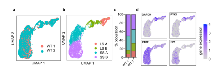
scRNA-seq identifies transcriptionally distinct long slender and short stumpy form T. brucei. To model stumpy differentiation in vitro the authors used a pleomorphic line, and began by treating parasites with oligopeptide-rich BHI broth, which can induce T. brucei bloodstream form differentiation in a titratable manner. To capture the transcriptomes of slender, intermediate and stumpy forms, they combined parasites after 0, 24, 48, or 72h after 10% BHI treatment in equal numbers. scRNA-Seq was then performed using the Chromium Single Cell 3’ workflow (10x genomics) and Illumina Sequencing, of two independent biological replicates with a total of 9344 cells examined. Medians of 1051 and 1439 genes were detected per cell. Cells from both experiments were integrated and visualized using UMAP. The authors identified four distinct groups containing transcriptionally similar cells, with two of those groups being clear slender and stumpy-like cells. The four groups were termed slender A, slender B, stumpy A and stumpy B. Differential expression analysis of the transcripts between the 4 groups showed significant overlap between the genes of group slender A and B, and between stumpy A and B. However, the study showed 183 markers unique to the slender A group, 95 to slender B, 55 to stumpy A, and 9 to stumpy B. Gene ontology enrichment analysis revealed the association of each cluster’s marker genes with distinct biological processes. Altogether, the authors emphasize that a distinct cluster representative of ‘the intermediate’ stage transcriptome between slender and stumpy forms was not evident.
Trajectory analysis of long slender to short stumpy differentiation. Given the overlap detected in the clustering analysis, the authors conducted trajectory interference and pseudotime analysis to study gene expression changes during stumpy development in detail. Here, individual cells were re-plotted as a PHATE (potential of heat-diffusion for affinity-based transition embedding) map, which allows preservation of the continual progression of developmental processes. They found that slender A and B clusters remained separate, while stumpy A and stumpy B showed significant overlap. This allowed identification of 2001 genes differentially expressed as a function of pseudotime, which were grouped into 9 modules of co-expressed genes showing similar patterns of expression throughout differentiation. 66% of those genes had been previously shown to be significantly differentially expressed between slender and stumpy populations isolated from low and peak parasitemias in vivo. Altogether, the authors highlight the advantage of scRNA-Seq to reveal transient events in an asynchronous developmental trajectory. Moreover, GO term enrichment for biological processes associated with each gene module revealed a potential order of biological events during slender-to-stumpy development. Besides of the annotated genes, 635 hypothetical genes were identified as differentially expressed during slender to stumpy differentiation. Altogether, pseudotime analysis allowed identification of novel genes differentially expressed during bloodstream form differentiation, as well as each gene’s detailed expression pattern.
Transcript abundance during the bloodstream slender cell cycle. Given that replicating slender bloodstream form cells were captured in the experiments described above, the authors went on to explore if the scRNA-Seq data could reveal greater detail than what is known, on gene expression changes during the cell cycle. Each cell was assigned to a cell cycle phase using marker genes previously identified in bulk RNA-Seq analyses. Slender A and B cells were grouped closer to cells of the same phase, with parasites most distal to Stumpy A and B labelled as late G1, followed by S and G2/M phase cells. Slender B cells most proximal to stumpy A contained all 4 cycle phases, although early G1 cells were enriched here. Interestingly, stumpy A and B cells were marked in a variety of cell cycle phases. An important finding of this section of the work was a) the identification of genes driving the cell cycle, and b) the identification of 3 genes previously shown to be involved in stumpy development with differential expression patterns in slender cells- namely, RBP7B (increased in late G1 cells through to G2/M), PPC2 (decreased in late G1/S phase parasites), and ZC3H20 (dropped in expression in late G1/S phase).
ZC3H20 null parasites fail to differentiate in response to BHI. ZC3H20 peaks in expression at the slender B to stumpy transition in pseudotime, and it has been previously shown to be required for differentiation in vivo and in vitro. Based on this, the authors used a ZC3H20 null T. brucei line to investigate where parasites fail in their development to stumpy forms with respect to transcriptome changes, and aimed to identify mRNA targets of ZC3H20 itself. Incubation of ZC3H20 KO in 10% BHI broth showed that these parasites continued to replicate beyond time points where WT cells had arrested, and after 72h of culture, failed to express PAD1. Moreover, consistent with their inability to produce stumpy forms, ZC3H20 KO failed to differentiate into procyclic cells. scRNA-Seq was then performed on ZC3H20 KO cells at 0, 24, 48, or 72h after 10% BHI treatment- as done for WT cells. Clustering the ZC3H20 KO and WT integrated cells resulted in 6 distinct clusters: stumpy A and B, and 4 slender clusters, called slender A.1, A.2, B.1, and B.2. While 77.3% of WT cells were found in clusters stumpy A or B, only 0.3% of ZC3H20 KO cells were in either, consistent with the near complete ablation of stumpy formation in the mutant parasites. The B.2 cohort was comprised almost entirely of ZC3H20 KO cells. Marker gene analysis between clusters identified 94 marker genes upregulated in slender B.2 cells, 18 of which were unique to this cluster.
Trajectory comparison between WT and ZC3H20 KO cells reveals functional separation of downregulation and upregulation of transcripts during differentiation. The authors then compared transcriptomic changes in ZC3H20 KO and WT parasites after BHI treatment, by inferring a trajectory from the WT and ZC3H20 KO integrated parasites. This identified a branched trajectory – while early in pseudotime WT and ZC3H20 KO parasites are transcriptionally similar and arrange on the same lineage, later there was a clear branching in their development, whereby WT cells ended in stumpy forms, and ZC3H20 KO in slender B.2. 587 genes of the 2001 identified as differentially expressed during stumpy development in WT cells, significantly changed in expression in ZC3H20 KO cells across the truncated trajectory. ZC3H20 KO cells failed to upregulate transcripts later in development that are required for stumpy formation, and this point of dysregulation coincided with the peak of ZC3H20 expression during normal WT differentiation.
The authors then aimed to identify regulators of early stumpy development, and so looked for genes which changed significantly in abundance at the start of the trajectory to a point downstream of the ZC3H20 branch. 234 genes changed in transcript abundance between these points, and were associated with trajectory progression. RDK2 and PAD2 were associated with both trajectories, but had different patterns of expression. 83 genes were differentially expressed only in the trajectory of WT parasites. Conversely, 35 genes with early altered expression were associated with the truncated ZC3H20 KO development only. Altogether, comparison of the differentiation of WT and ZC3H20 KO cells through scRNA-Seq allowed the identification of direct and indirect targets of ZC3H20 altered specifically during differentiation; the failure point of ZC3H20 KO cells during differentiation; and putative immediate early regulators of differentiation.
What I like about this preprint
I think the question addressed in this work is one that remained outstanding in the field of T. brucei, namely, little is known about the intermediate stages of development of the parasite from slender to stumpy forms. I think the use of scRNA-Seq in T. brucei research, and in this work allowed opening various avenues of research, relevant to the whole field.
References
- Briggs et al, Single cell transcriptomic analysis of bloodstream form T. brucei reconstructs cell cycle progression and differentiation via quorum sensing. bioRxiv, 2020.
doi: https://doi.org/10.1242/prelights.26638
Read preprint (No Ratings Yet)
Sign up to customise the site to your preferences and to receive alerts
Register hereAlso in the cell biology category:
Combinatorial and Inducible CRISPRa/i Enables Canalized hiPSC Forward Programming and Iterative Refinement via Single-Cell Genomics
Cell-ID
Developmental conversion of the nucleolus into an RNA Polymerase II transcriptional platform in Drosophila spermatocytes
Panagiotis Giannios
Cell position is more important than cell shape or age for the acquisition of cell identity in the brown alga Ectocarpus
Urvashi Goswami
preLists in the cell biology category:
Developmental regulation: molecular and ecological niches
This conference was held at the Station Biologique de Roscoff (France) and brought together researchers exploring how diverse niche environments shape developmental processes across scales. Spanning topics from ecological and metabolic influences to signalling networks, mechanics and gene regulation, the meeting highlighted the interplay between intrinsic and extrinsic factors in controlling cell fate and tissue organisation. This preList gathers preprints discussed by speakers and poster presenters during the meeting. Please do get in touch at preLights@biologists.com if you notice any relevant preprints that we may have missed.
| List by | Ingrid Tsang |
preLighters’ choice – Handpicked DevBio preprints
preLighters with expertise across developmental and stem cell biology have nominated a few developmental biology (and related) preprints they’re excited about and explain in a few paragraph why. Concise preprint highlights, prepared by the preLighter community – a quick way to spot upcoming trends, new methods and fresh ideas.
| List by | Theodora Stougiannou et al. |
BSDB Spring Meeting: Molecules to Morphogenesis
The British Society for Developmental Biology (BSDB) Spring Meeting Molecules to Morphogenesis was held from 23–26 March 2026 at the University of Warwick (UK). This meeting brought together a vibrant community of researchers to discuss how molecular mechanisms are integrated across scales to drive morphogenesis, spanning diverse model systems and approaches. This preList contains preprints by presenters from the talk and poster sessions at the meeting. Please do get in touch at preLights@biologists.com if you notice any relevant preprints that we may have missed.
| List by | Ingrid Tsang |
Keystone Symposium on Stem Cell Models in Embryology 2026
The Keystone Symposium on Stem Cell Models in Embryology, 2026, was organised by Jun Wu (UT Southwestern), Jianping Fu (University of Michigan) and Miki Ebisuya (TU Dresden) and held at Asilomar Conference Grounds in California (US). The meeting discussed recent advances made in establishing stem-cell-based embryo models, what fundamental insights into developmental processes have been gleaned from them, as well as how they are beginning to be applied more widely. This prelist contains preprints by presenters at the talk and poster sessions at the conference, which our Reviews Editor in attendance spotted. Please do reach out to preLights@biologists.com if you notice any that we’ve missed.
| List by | Ingrid Tsang |
SciELO preprints – From 2025 onwards
SciELO has become a cornerstone of open, multilingual scholarly communication across Latin America. Its preprint server, SciELO preprints, is expanding the global reach of preprinted research from the region (for more information, see our interview with Carolina Tanigushi). This preList brings together biological, English language SciELO preprints to help readers discover emerging work from the Global South. By highlighting these preprints in one place, we aim to support visibility, encourage early feedback, and showcase the vibrant research communities contributing to SciELO’s open science ecosystem.
| List by | Carolina Tanigushi |
November in preprints – DevBio & Stem cell biology
preLighters with expertise across developmental and stem cell biology have nominated a few developmental and stem cell biology (and related) preprints posted in November they’re excited about and explain in a single paragraph why. Concise preprint highlights, prepared by the preLighter community – a quick way to spot upcoming trends, new methods and fresh ideas.
| List by | Aline Grata et al. |
October in preprints – DevBio & Stem cell biology
Each month, preLighters with expertise across developmental and stem cell biology nominate a few recent developmental and stem cell biology (and related) preprints they’re excited about and explain in a single paragraph why. Short, snappy picks from working scientists — a quick way to spot fresh ideas, bold methods and papers worth reading in full. These preprints can all be found in the October preprint list published on the Node.
| List by | Deevitha Balasubramanian et al. |
October in preprints – Cell biology edition
Different preLighters, with expertise across cell biology, have worked together to create this preprint reading list for researchers with an interest in cell biology. This month, most picks fall under (1) Cell organelles and organisation, followed by (2) Mechanosignaling and mechanotransduction, (3) Cell cycle and division and (4) Cell migration
| List by | Matthew Davies et al. |
September in preprints – Cell biology edition
A group of preLighters, with expertise in different areas of cell biology, have worked together to create this preprint reading list. This month, categories include: (1) Cell organelles and organisation, (2) Cell signalling and mechanosensing, (3) Cell metabolism, (4) Cell cycle and division, (5) Cell migration
| List by | Sristilekha Nath et al. |
July in preprints – the CellBio edition
A group of preLighters, with expertise in different areas of cell biology, have worked together to create this preprint reading lists for researchers with an interest in cell biology. This month, categories include: (1) Cell Signalling and Mechanosensing (2) Cell Cycle and Division (3) Cell Migration and Cytoskeleton (4) Cancer Biology (5) Cell Organelles and Organisation
| List by | Girish Kale et al. |
June in preprints – the CellBio edition
A group of preLighters, with expertise in different areas of cell biology, have worked together to create this preprint reading lists for researchers with an interest in cell biology. This month, categories include: (1) Cell organelles and organisation (2) Cell signaling and mechanosensation (3) Genetics/gene expression (4) Biochemistry (5) Cytoskeleton
| List by | Barbora Knotkova et al. |
May in preprints – the CellBio edition
A group of preLighters, with expertise in different areas of cell biology, have worked together to create this preprint reading lists for researchers with an interest in cell biology. This month, categories include: 1) Biochemistry/metabolism 2) Cancer cell Biology 3) Cell adhesion, migration and cytoskeleton 4) Cell organelles and organisation 5) Cell signalling and 6) Genetics
| List by | Barbora Knotkova et al. |
Keystone Symposium – Metabolic and Nutritional Control of Development and Cell Fate
This preList contains preprints discussed during the Metabolic and Nutritional Control of Development and Cell Fate Keystone Symposia. This conference was organized by Lydia Finley and Ralph J. DeBerardinis and held in the Wylie Center and Tupper Manor at Endicott College, Beverly, MA, United States from May 7th to 9th 2025. This meeting marked the first in-person gathering of leading researchers exploring how metabolism influences development, including processes like cell fate, tissue patterning, and organ function, through nutrient availability and metabolic regulation. By integrating modern metabolic tools with genetic and epidemiological insights across model organisms, this event highlighted key mechanisms and identified open questions to advance the emerging field of developmental metabolism.
| List by | Virginia Savy, Martin Estermann |
April in preprints – the CellBio edition
A group of preLighters, with expertise in different areas of cell biology, have worked together to create this preprint reading lists for researchers with an interest in cell biology. This month, categories include: 1) biochemistry/metabolism 2) cell cycle and division 3) cell organelles and organisation 4) cell signalling and mechanosensing 5) (epi)genetics
| List by | Vibha SINGH et al. |
March in preprints – the CellBio edition
A group of preLighters, with expertise in different areas of cell biology, have worked together to create this preprint reading lists for researchers with an interest in cell biology. This month, categories include: 1) cancer biology 2) cell migration 3) cell organelles and organisation 4) cell signalling and mechanosensing 5) genetics and genomics 6) other
| List by | Girish Kale et al. |
Biologists @ 100 conference preList
This preList aims to capture all preprints being discussed at the Biologists @100 conference in Liverpool, UK, either as part of the poster sessions or the (flash/short/full-length) talks.
| List by | Reinier Prosee, Jonathan Townson |
February in preprints – the CellBio edition
A group of preLighters, with expertise in different areas of cell biology, have worked together to create this preprint reading lists for researchers with an interest in cell biology. This month, categories include: 1) biochemistry and cell metabolism 2) cell organelles and organisation 3) cell signalling, migration and mechanosensing
| List by | Barbora Knotkova et al. |
Community-driven preList – Immunology
In this community-driven preList, a group of preLighters, with expertise in different areas of immunology have worked together to create this preprint reading list.
| List by | Felipe Del Valle Batalla et al. |
January in preprints – the CellBio edition
A group of preLighters, with expertise in different areas of cell biology, have worked together to create this preprint reading lists for researchers with an interest in cell biology. This month, categories include: 1) biochemistry/metabolism 2) cell migration 3) cell organelles and organisation 4) cell signalling and mechanosensing 5) genetics/gene expression
| List by | Barbora Knotkova et al. |
December in preprints – the CellBio edition
A group of preLighters, with expertise in different areas of cell biology, have worked together to create this preprint reading lists for researchers with an interest in cell biology. This month, categories include: 1) cell cycle and division 2) cell migration and cytoskeleton 3) cell organelles and organisation 4) cell signalling and mechanosensing 5) genetics/gene expression
| List by | Matthew Davies et al. |
November in preprints – the CellBio edition
This is the first community-driven preList! A group of preLighters, with expertise in different areas of cell biology, have worked together to create this preprint reading lists for researchers with an interest in cell biology. Categories include: 1) cancer cell biology 2) cell cycle and division 3) cell migration and cytoskeleton 4) cell organelles and organisation 5) cell signalling and mechanosensing 6) genetics/gene expression
| List by | Felipe Del Valle Batalla et al. |
BSCB-Biochemical Society 2024 Cell Migration meeting
This preList features preprints that were discussed and presented during the BSCB-Biochemical Society 2024 Cell Migration meeting in Birmingham, UK in April 2024. Kindly put together by Sara Morais da Silva, Reviews Editor at Journal of Cell Science.
| List by | Reinier Prosee |
‘In preprints’ from Development 2022-2023
A list of the preprints featured in Development's 'In preprints' articles between 2022-2023
| List by | Alex Eve, Katherine Brown |
preLights peer support – preprints of interest
This is a preprint repository to organise the preprints and preLights covered through the 'preLights peer support' initiative.
| List by | preLights peer support |
The Society for Developmental Biology 82nd Annual Meeting
This preList is made up of the preprints discussed during the Society for Developmental Biology 82nd Annual Meeting that took place in Chicago in July 2023.
| List by | Joyce Yu, Katherine Brown |
CSHL 87th Symposium: Stem Cells
Preprints mentioned by speakers at the #CSHLsymp23
| List by | Alex Eve |
Journal of Cell Science meeting ‘Imaging Cell Dynamics’
This preList highlights the preprints discussed at the JCS meeting 'Imaging Cell Dynamics'. The meeting was held from 14 - 17 May 2023 in Lisbon, Portugal and was organised by Erika Holzbaur, Jennifer Lippincott-Schwartz, Rob Parton and Michael Way.
| List by | Helen Zenner |
9th International Symposium on the Biology of Vertebrate Sex Determination
This preList contains preprints discussed during the 9th International Symposium on the Biology of Vertebrate Sex Determination. This conference was held in Kona, Hawaii from April 17th to 21st 2023.
| List by | Martin Estermann |
Alumni picks – preLights 5th Birthday
This preList contains preprints that were picked and highlighted by preLights Alumni - an initiative that was set up to mark preLights 5th birthday. More entries will follow throughout February and March 2023.
| List by | Sergio Menchero et al. |
CellBio 2022 – An ASCB/EMBO Meeting
This preLists features preprints that were discussed and presented during the CellBio 2022 meeting in Washington, DC in December 2022.
| List by | Nadja Hümpfer et al. |
Fibroblasts
The advances in fibroblast biology preList explores the recent discoveries and preprints of the fibroblast world. Get ready to immerse yourself with this list created for fibroblasts aficionados and lovers, and beyond. Here, my goal is to include preprints of fibroblast biology, heterogeneity, fate, extracellular matrix, behavior, topography, single-cell atlases, spatial transcriptomics, and their matrix!
| List by | Osvaldo Contreras |
EMBL Synthetic Morphogenesis: From Gene Circuits to Tissue Architecture (2021)
A list of preprints mentioned at the #EESmorphoG virtual meeting in 2021.
| List by | Alex Eve |
FENS 2020
A collection of preprints presented during the virtual meeting of the Federation of European Neuroscience Societies (FENS) in 2020
| List by | Ana Dorrego-Rivas |
Planar Cell Polarity – PCP
This preList contains preprints about the latest findings on Planar Cell Polarity (PCP) in various model organisms at the molecular, cellular and tissue levels.
| List by | Ana Dorrego-Rivas |
BioMalPar XVI: Biology and Pathology of the Malaria Parasite
[under construction] Preprints presented at the (fully virtual) EMBL BioMalPar XVI, 17-18 May 2020 #emblmalaria
| List by | Dey Lab, Samantha Seah |
1
Cell Polarity
Recent research from the field of cell polarity is summarized in this list of preprints. It comprises of studies focusing on various forms of cell polarity ranging from epithelial polarity, planar cell polarity to front-to-rear polarity.
| List by | Yamini Ravichandran |
TAGC 2020
Preprints recently presented at the virtual Allied Genetics Conference, April 22-26, 2020. #TAGC20
| List by | Maiko Kitaoka et al. |
3D Gastruloids
A curated list of preprints related to Gastruloids (in vitro models of early development obtained by 3D aggregation of embryonic cells). Updated until July 2021.
| List by | Paul Gerald L. Sanchez and Stefano Vianello |
ECFG15 – Fungal biology
Preprints presented at 15th European Conference on Fungal Genetics 17-20 February 2020 Rome
| List by | Hiral Shah |
ASCB EMBO Annual Meeting 2019
A collection of preprints presented at the 2019 ASCB EMBO Meeting in Washington, DC (December 7-11)
| List by | Madhuja Samaddar et al. |
EMBL Seeing is Believing – Imaging the Molecular Processes of Life
Preprints discussed at the 2019 edition of Seeing is Believing, at EMBL Heidelberg from the 9th-12th October 2019
| List by | Dey Lab |
Autophagy
Preprints on autophagy and lysosomal degradation and its role in neurodegeneration and disease. Includes molecular mechanisms, upstream signalling and regulation as well as studies on pharmaceutical interventions to upregulate the process.
| List by | Sandra Malmgren Hill |
Lung Disease and Regeneration
This preprint list compiles highlights from the field of lung biology.
| List by | Rob Hynds |
Cellular metabolism
A curated list of preprints related to cellular metabolism at Biorxiv by Pablo Ranea Robles from the Prelights community. Special interest on lipid metabolism, peroxisomes and mitochondria.
| List by | Pablo Ranea Robles |
BSCB/BSDB Annual Meeting 2019
Preprints presented at the BSCB/BSDB Annual Meeting 2019
| List by | Dey Lab |
MitoList
This list of preprints is focused on work expanding our knowledge on mitochondria in any organism, tissue or cell type, from the normal biology to the pathology.
| List by | Sandra Franco Iborra |
Biophysical Society Annual Meeting 2019
Few of the preprints that were discussed in the recent BPS annual meeting at Baltimore, USA
| List by | Joseph Jose Thottacherry |
ASCB/EMBO Annual Meeting 2018
This list relates to preprints that were discussed at the recent ASCB conference.
| List by | Dey Lab, Amanda Haage |