


PP1 and PP2A use opposite phospho-dependencies to control distinct processes at the kinetochore
Posted on: 17 July 2019 , updated on: 18 July 2019
Preprint posted on 5 May 2019
Article now published in Cell Reports at http://dx.doi.org/10.1016/j.celrep.2019.07.067
How do highly similar phosphatases achieve functional specificity? The motifs recruiting them reveal more than you’d expect!
Selected by Angika BasantCategories: cell biology
Background:
There are about 400 Ser/Thr kinases in the human genome but only 28 known Ser/Thr phosphatases (1, 2). How does this small group of phosphatases accurately regulate the phospho-state of hundreds of substrates?
PP1 and PP2A-B56 belong to the same PPP family of phosphatases. They share a similar 280 residue catalytic domain, the activities of which are comparable (3). Yet one does not appear to fully compensate the loss of the other in vivo (4), suggesting that each performs many unique functions. Why and how is a given phosphatase better-suited for certain roles over another? What features make it distinct?
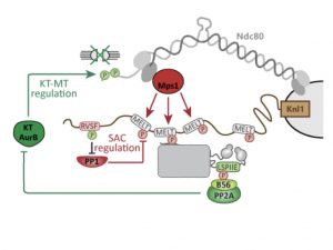
This study addresses these questions in the context of the kinetochore. During spindle assembly, the kinetochore needs to perform two critical functions. It must (A) make stable, accurate attachments with microtubules and (B) signal the cell to maintain mitotic state until (A) has been achieved. These two functions are controlled by the phospho-states of the kinetochore protein Ndc80 and MELT motifs of the Knl1 protein respectively. Aurora B kinase phosphorylation of Ndc80 destabilises incorrect kinetochore-microtubule interactions. MELT motif phosphorylation by Mps1 kinase signals the cell to remain in mitosis. These two key phospho-modifications are also regulated by kinetochore phosphatases.
Both PP1 and PP2A-B56 are recruited to the Knl1 subcomplex on the kinetochore. PP1 directly binds the N-terminus of Knl1 to a RVSF motif. PP2A-B56 binds a LSPIIE motif of a Knl1 interactor. Interestingly, both these motifs are phosphorylated. And rather curiously, this has opposite effects on binding of the respective phosphatase. PP1 binding is reduced by phosphorylation of RVSF (5, 6), while PP2A-B56 binding is enhanced by LSPIIE phosphorylation (7, 8, 9).
Why are two similar phosphatases recruited to the same scaffold? Do they perform different functions? How are their individual activities regulated? Do their dissimilar binding patterns play a role?
Key findings:
The authors first establish that the PP1 and PP2A-B56 do not perform identical functions at the kinetochore. Deleting LSPIIE, but not RVSF, results in increased Ndc80 phosphorylation and chromosome alignment defects. This implies that in a wild-type kinetochore, only PP2A-B56 directly regulates phospho-Ndc80.
Removal of either LSPIIE or RVSF results in mitotic arrest and high phospho-MELT. However, both phosphatases do not directly dephosphorylate MELT. PP2A-B56 is known to promote PP1 association to the kinetochore by dephosphorylating Knl1 (10). When these Knl1 phosphosites are mutated, removal of the LSPIIE motif no longer causes mitotic arrest, while deleting RVSF still results in arrest and high phospho-MELT. Collectively, these results indicate that only PP1 directly regulates mitotic arrest/exit. While PP2A-B56 indirectly facilitates PP1 recruitment, it does not dephosphorylate MELT motifs on wild-type kinetochores (even though it is detectable there).
Two hypotheses were tested to explain these differences. First – the identity of the phosphatase could be critical to each function. However, deleting the motif recruiting one and artificially recruiting the other in its place had no measurable effects.
Second, the authors considered whether the exact binding position of the phosphatase matters to function by say, controlling substrate accessibility. But deleting the motif recruiting either phosphatase and recruiting the same phosphatase to a different site on Knl1 did not impact its function. Ndc80 dephosphorylation occurred normally when PP2A-B56 was repositioned and MELT dephosphorylation was not impacted by PP1 repositioning.
If the two enzymes can functionally substitute for each other and their positions within the Knl1 subcomplex is immaterial to signalling, how are PP1 and PP2A-B56 restricted to distinct functions at the kinetochore?
As mentioned previously, a key difference between the two enzymes is the mechanism by which they are recruited to their respective motifs. To explore this aspect, the authors focused on a critical unexplained observation that despite being present at the kinetochore, PP2A-B56 does not dephosphorylate MELT motifs in the absence of PP1. They take advantage of a motif LPTIHE which binds PP2A-B56 with an affinity similar to LSPIIE but independently of phosphorylation. Amazingly, in the absence of RVSF (and thereby PP1), if the motif recruiting PP2A-B56 is changed to LPTIHE, PP2A-B56 is now able to substitute for PP1 and induce cells to exit mitosis.
The authors next construct a mathematical model for this network. It assumes that kinase activities involved in the system are fixed and that both PP1 and PP2A-B56 dephosphorylate the following substrates with the same kinetics, namely MELT, Ndc80, RVSP and LSPIIE. The only difference is that PP2A-B56 is recruited by phosphorylation of the motif it binds to, and PP1 is not. Strikingly, such a model is able to recapitulate their experimental findings.
Finally, in a wider search for such motifs, the authors find that RVxF and LxxIxE are present in ~700 proteins and a large fraction of these contain phosphorylatable residues in key positions. Quite interestingly, structural examination of PP1 and B56 reveals how a negatively charged surface on the former and a basic groove on B56 would explain the opposite behaviour towards a negatively charged phosphate in the motifs they bind.
What I like about this preprint:
This paper tackles a complex but widely-applicable problem with a nice combination of clear experiments, mathematical modelling and a wider analysis of the implicated motifs in other contexts. The key finding of this paper that inverse phospho-dependencies in phosphatase binding can create unique functional differences between similar enzymes, is unexpected and novel.
Questions for the authors:
- How critical are binding kinetics to the manifestation of these feedback loops? For example, if FRAP recovery of PP2A-B56 accumulation at the kinetochore was compared between LSPIIE- and LPTIHE-driven binding, would they be significantly different? And does that matter for the striking result seen in Figure 4E?
- Among the validated proteins with RVxF/LxxIxE motifs that are phosphorylated, are the predicted binding partners more often kinases and phosphatases? That is, is such phospho-dependence more likely to impact a certain type of signalling network and do you predict that tyrosine kinases/phosphatases may be regulated this way, albeit with different motifs?
References:
- Almo et al., Struct. Funct. Genomics, 2007
- Manning et al., Science, 2002
- Ingebritsen and Cohen, Eur J Biochem, 1983
- Kauko et al., bioRxiv, 2018
- Kim et al., J Bio Chem, 2003
- Nasa et al., Sci Signal, 2018
- Wang et al., Protein Cell, 2016
- Wang et al., Structure, 2016
- Hertz et al., Mol Cell, 2016
- Nijenhuis et al., Nat Cell Biol, 2014
doi: https://doi.org/10.1242/prelights.12195
Read preprint (3 votes)
 Sign up to customise the site to your preferences and to receive alerts
Register hereAlso in the cell biology category:
Resilience to cardiac aging in Greenland shark Somniosus microcephalus
Theodora Stougiannou
The lipidomic architecture of the mouse brain
CRM UoE Journal Club et al.
Self-renewal of neuronal mitochondria through asymmetric division
Lorena Olifiers
preLists in the cell biology category:
SciELO preprints – From 2025 onwards
SciELO has become a cornerstone of open, multilingual scholarly communication across Latin America. Its preprint server, SciELO preprints, is expanding the global reach of preprinted research from the region (for more information, see our interview with Carolina Tanigushi). This preList brings together biological, English language SciELO preprints to help readers discover emerging work from the Global South. By highlighting these preprints in one place, we aim to support visibility, encourage early feedback, and showcase the vibrant research communities contributing to SciELO’s open science ecosystem.
| List by | Carolina Tanigushi |
November in preprints – DevBio & Stem cell biology
preLighters with expertise across developmental and stem cell biology have nominated a few developmental and stem cell biology (and related) preprints posted in November they’re excited about and explain in a single paragraph why. Concise preprint highlights, prepared by the preLighter community – a quick way to spot upcoming trends, new methods and fresh ideas.
| List by | Aline Grata et al. |
October in preprints – DevBio & Stem cell biology
Each month, preLighters with expertise across developmental and stem cell biology nominate a few recent developmental and stem cell biology (and related) preprints they’re excited about and explain in a single paragraph why. Short, snappy picks from working scientists — a quick way to spot fresh ideas, bold methods and papers worth reading in full. These preprints can all be found in the October preprint list published on the Node.
| List by | Deevitha Balasubramanian et al. |
October in preprints – Cell biology edition
Different preLighters, with expertise across cell biology, have worked together to create this preprint reading list for researchers with an interest in cell biology. This month, most picks fall under (1) Cell organelles and organisation, followed by (2) Mechanosignaling and mechanotransduction, (3) Cell cycle and division and (4) Cell migration
| List by | Matthew Davies et al. |
September in preprints – Cell biology edition
A group of preLighters, with expertise in different areas of cell biology, have worked together to create this preprint reading list. This month, categories include: (1) Cell organelles and organisation, (2) Cell signalling and mechanosensing, (3) Cell metabolism, (4) Cell cycle and division, (5) Cell migration
| List by | Sristilekha Nath et al. |
July in preprints – the CellBio edition
A group of preLighters, with expertise in different areas of cell biology, have worked together to create this preprint reading lists for researchers with an interest in cell biology. This month, categories include: (1) Cell Signalling and Mechanosensing (2) Cell Cycle and Division (3) Cell Migration and Cytoskeleton (4) Cancer Biology (5) Cell Organelles and Organisation
| List by | Girish Kale et al. |
June in preprints – the CellBio edition
A group of preLighters, with expertise in different areas of cell biology, have worked together to create this preprint reading lists for researchers with an interest in cell biology. This month, categories include: (1) Cell organelles and organisation (2) Cell signaling and mechanosensation (3) Genetics/gene expression (4) Biochemistry (5) Cytoskeleton
| List by | Barbora Knotkova et al. |
May in preprints – the CellBio edition
A group of preLighters, with expertise in different areas of cell biology, have worked together to create this preprint reading lists for researchers with an interest in cell biology. This month, categories include: 1) Biochemistry/metabolism 2) Cancer cell Biology 3) Cell adhesion, migration and cytoskeleton 4) Cell organelles and organisation 5) Cell signalling and 6) Genetics
| List by | Barbora Knotkova et al. |
Keystone Symposium – Metabolic and Nutritional Control of Development and Cell Fate
This preList contains preprints discussed during the Metabolic and Nutritional Control of Development and Cell Fate Keystone Symposia. This conference was organized by Lydia Finley and Ralph J. DeBerardinis and held in the Wylie Center and Tupper Manor at Endicott College, Beverly, MA, United States from May 7th to 9th 2025. This meeting marked the first in-person gathering of leading researchers exploring how metabolism influences development, including processes like cell fate, tissue patterning, and organ function, through nutrient availability and metabolic regulation. By integrating modern metabolic tools with genetic and epidemiological insights across model organisms, this event highlighted key mechanisms and identified open questions to advance the emerging field of developmental metabolism.
| List by | Virginia Savy, Martin Estermann |
April in preprints – the CellBio edition
A group of preLighters, with expertise in different areas of cell biology, have worked together to create this preprint reading lists for researchers with an interest in cell biology. This month, categories include: 1) biochemistry/metabolism 2) cell cycle and division 3) cell organelles and organisation 4) cell signalling and mechanosensing 5) (epi)genetics
| List by | Vibha SINGH et al. |
March in preprints – the CellBio edition
A group of preLighters, with expertise in different areas of cell biology, have worked together to create this preprint reading lists for researchers with an interest in cell biology. This month, categories include: 1) cancer biology 2) cell migration 3) cell organelles and organisation 4) cell signalling and mechanosensing 5) genetics and genomics 6) other
| List by | Girish Kale et al. |
Biologists @ 100 conference preList
This preList aims to capture all preprints being discussed at the Biologists @100 conference in Liverpool, UK, either as part of the poster sessions or the (flash/short/full-length) talks.
| List by | Reinier Prosee, Jonathan Townson |
February in preprints – the CellBio edition
A group of preLighters, with expertise in different areas of cell biology, have worked together to create this preprint reading lists for researchers with an interest in cell biology. This month, categories include: 1) biochemistry and cell metabolism 2) cell organelles and organisation 3) cell signalling, migration and mechanosensing
| List by | Barbora Knotkova et al. |
Community-driven preList – Immunology
In this community-driven preList, a group of preLighters, with expertise in different areas of immunology have worked together to create this preprint reading list.
| List by | Felipe Del Valle Batalla et al. |
January in preprints – the CellBio edition
A group of preLighters, with expertise in different areas of cell biology, have worked together to create this preprint reading lists for researchers with an interest in cell biology. This month, categories include: 1) biochemistry/metabolism 2) cell migration 3) cell organelles and organisation 4) cell signalling and mechanosensing 5) genetics/gene expression
| List by | Barbora Knotkova et al. |
December in preprints – the CellBio edition
A group of preLighters, with expertise in different areas of cell biology, have worked together to create this preprint reading lists for researchers with an interest in cell biology. This month, categories include: 1) cell cycle and division 2) cell migration and cytoskeleton 3) cell organelles and organisation 4) cell signalling and mechanosensing 5) genetics/gene expression
| List by | Matthew Davies et al. |
November in preprints – the CellBio edition
This is the first community-driven preList! A group of preLighters, with expertise in different areas of cell biology, have worked together to create this preprint reading lists for researchers with an interest in cell biology. Categories include: 1) cancer cell biology 2) cell cycle and division 3) cell migration and cytoskeleton 4) cell organelles and organisation 5) cell signalling and mechanosensing 6) genetics/gene expression
| List by | Felipe Del Valle Batalla et al. |
BSCB-Biochemical Society 2024 Cell Migration meeting
This preList features preprints that were discussed and presented during the BSCB-Biochemical Society 2024 Cell Migration meeting in Birmingham, UK in April 2024. Kindly put together by Sara Morais da Silva, Reviews Editor at Journal of Cell Science.
| List by | Reinier Prosee |
‘In preprints’ from Development 2022-2023
A list of the preprints featured in Development's 'In preprints' articles between 2022-2023
| List by | Alex Eve, Katherine Brown |
preLights peer support – preprints of interest
This is a preprint repository to organise the preprints and preLights covered through the 'preLights peer support' initiative.
| List by | preLights peer support |
The Society for Developmental Biology 82nd Annual Meeting
This preList is made up of the preprints discussed during the Society for Developmental Biology 82nd Annual Meeting that took place in Chicago in July 2023.
| List by | Joyce Yu, Katherine Brown |
CSHL 87th Symposium: Stem Cells
Preprints mentioned by speakers at the #CSHLsymp23
| List by | Alex Eve |
Journal of Cell Science meeting ‘Imaging Cell Dynamics’
This preList highlights the preprints discussed at the JCS meeting 'Imaging Cell Dynamics'. The meeting was held from 14 - 17 May 2023 in Lisbon, Portugal and was organised by Erika Holzbaur, Jennifer Lippincott-Schwartz, Rob Parton and Michael Way.
| List by | Helen Zenner |
9th International Symposium on the Biology of Vertebrate Sex Determination
This preList contains preprints discussed during the 9th International Symposium on the Biology of Vertebrate Sex Determination. This conference was held in Kona, Hawaii from April 17th to 21st 2023.
| List by | Martin Estermann |
Alumni picks – preLights 5th Birthday
This preList contains preprints that were picked and highlighted by preLights Alumni - an initiative that was set up to mark preLights 5th birthday. More entries will follow throughout February and March 2023.
| List by | Sergio Menchero et al. |
CellBio 2022 – An ASCB/EMBO Meeting
This preLists features preprints that were discussed and presented during the CellBio 2022 meeting in Washington, DC in December 2022.
| List by | Nadja Hümpfer et al. |
Fibroblasts
The advances in fibroblast biology preList explores the recent discoveries and preprints of the fibroblast world. Get ready to immerse yourself with this list created for fibroblasts aficionados and lovers, and beyond. Here, my goal is to include preprints of fibroblast biology, heterogeneity, fate, extracellular matrix, behavior, topography, single-cell atlases, spatial transcriptomics, and their matrix!
| List by | Osvaldo Contreras |
EMBL Synthetic Morphogenesis: From Gene Circuits to Tissue Architecture (2021)
A list of preprints mentioned at the #EESmorphoG virtual meeting in 2021.
| List by | Alex Eve |
FENS 2020
A collection of preprints presented during the virtual meeting of the Federation of European Neuroscience Societies (FENS) in 2020
| List by | Ana Dorrego-Rivas |
Planar Cell Polarity – PCP
This preList contains preprints about the latest findings on Planar Cell Polarity (PCP) in various model organisms at the molecular, cellular and tissue levels.
| List by | Ana Dorrego-Rivas |
BioMalPar XVI: Biology and Pathology of the Malaria Parasite
[under construction] Preprints presented at the (fully virtual) EMBL BioMalPar XVI, 17-18 May 2020 #emblmalaria
| List by | Dey Lab, Samantha Seah |
1
Cell Polarity
Recent research from the field of cell polarity is summarized in this list of preprints. It comprises of studies focusing on various forms of cell polarity ranging from epithelial polarity, planar cell polarity to front-to-rear polarity.
| List by | Yamini Ravichandran |
TAGC 2020
Preprints recently presented at the virtual Allied Genetics Conference, April 22-26, 2020. #TAGC20
| List by | Maiko Kitaoka et al. |
3D Gastruloids
A curated list of preprints related to Gastruloids (in vitro models of early development obtained by 3D aggregation of embryonic cells). Updated until July 2021.
| List by | Paul Gerald L. Sanchez and Stefano Vianello |
ECFG15 – Fungal biology
Preprints presented at 15th European Conference on Fungal Genetics 17-20 February 2020 Rome
| List by | Hiral Shah |
ASCB EMBO Annual Meeting 2019
A collection of preprints presented at the 2019 ASCB EMBO Meeting in Washington, DC (December 7-11)
| List by | Madhuja Samaddar et al. |
EMBL Seeing is Believing – Imaging the Molecular Processes of Life
Preprints discussed at the 2019 edition of Seeing is Believing, at EMBL Heidelberg from the 9th-12th October 2019
| List by | Dey Lab |
Autophagy
Preprints on autophagy and lysosomal degradation and its role in neurodegeneration and disease. Includes molecular mechanisms, upstream signalling and regulation as well as studies on pharmaceutical interventions to upregulate the process.
| List by | Sandra Malmgren Hill |
Lung Disease and Regeneration
This preprint list compiles highlights from the field of lung biology.
| List by | Rob Hynds |
Cellular metabolism
A curated list of preprints related to cellular metabolism at Biorxiv by Pablo Ranea Robles from the Prelights community. Special interest on lipid metabolism, peroxisomes and mitochondria.
| List by | Pablo Ranea Robles |
BSCB/BSDB Annual Meeting 2019
Preprints presented at the BSCB/BSDB Annual Meeting 2019
| List by | Dey Lab |
MitoList
This list of preprints is focused on work expanding our knowledge on mitochondria in any organism, tissue or cell type, from the normal biology to the pathology.
| List by | Sandra Franco Iborra |
Biophysical Society Annual Meeting 2019
Few of the preprints that were discussed in the recent BPS annual meeting at Baltimore, USA
| List by | Joseph Jose Thottacherry |
ASCB/EMBO Annual Meeting 2018
This list relates to preprints that were discussed at the recent ASCB conference.
| List by | Dey Lab, Amanda Haage |